Decoding the world’s deadliest animal
Illustration by Jasu Hu

Forefront

Decoding the world’s deadliest animal

Illustration by Jasu Hu

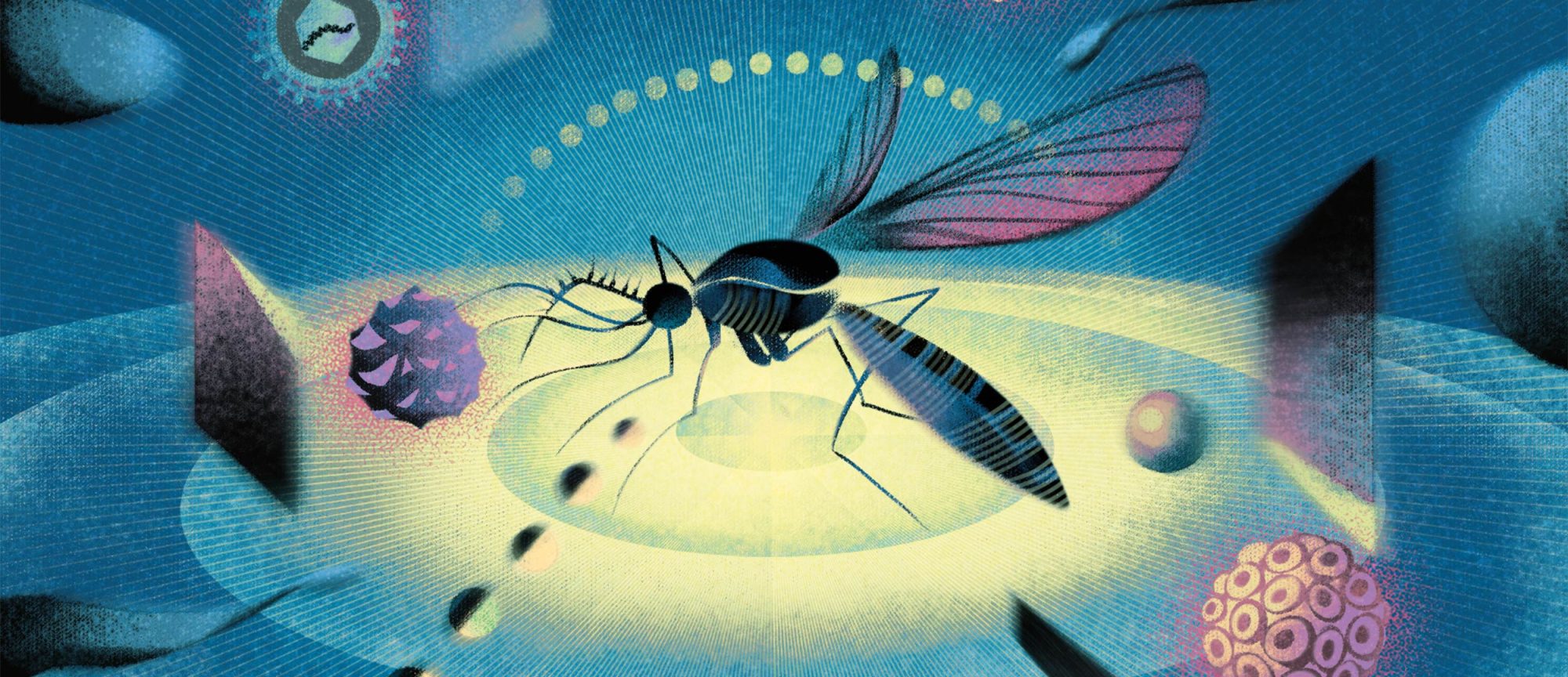
Mosquitoes aren’t just irritating: As the primary vectors for malaria, yellow fever, Zika, and a host of other dangerous viruses, these tiny creatures are by far the most dangerous animals on the planet.

But deep dives into mosquito genetics and behavior by Leslie B. Vosshall’s lab could change that. This year, Vosshall and her colleagues made two major advances: one that corrects a long- standing misconception about mosquito mating and another that reveals the insect’s genetic secrets, cell by cell.

In the first study, postdoc Leah Houri-Zeevi uncovered the first evidence of what happens when a female mosquito chooses to mate for the one and only time in her life: a subtle movement of her genitalia that allows insemination to occur. This places the female firmly in control of copulation—a finding that overturns the decades-old assumption that male mosquitoes run the show.

“It’s really profound that the field assumed for so long that the female must be passive,” Vosshall notes. “Sometimes you need to pick apart an accepted dogma to see if there’s actually evidence to back it up. In this case, there wasn’t.”

In addition to upending decades of conventional wisdom, the discovery also helps explain why some mosquito species are outcompeting others. It could ultimately lead to new ways of interfering with mosquito reproduction, driving down the numbers of these deadly disease vectors. (A single female can lay more than 1,000 eggs over her lifetime.)

Achieving this breakthrough was no mean feat: A mosquito mating lasts for only 14 seconds, and the phase that includes the team’s key finding takes only one or two.

But by combining high-speed cameras, artificial intelligence, and genetically engineered mosquitoes equipped with fluorescent sperm, Houri-Zeevi and her colleagues were ultimately able to determine what leads to a successful coupling: The male inserts structures called gonostyli into the female’s genital tip and vibrates them; if she wants to mate, she elongates her own genital tip, permitting the male to transfer his sperm.

“If she doesn’t make this movement, it doesn’t matter what the male does—no successful mating will occur,” says Houri-Zeevi. “And when previously mated females pair up with a male, no elongation happens. It’s a one-and-done experience for her.”

In the second study, Nadav Shai, a senior scientist in the Vosshall lab, led a global collaboration to create the first-ever cellular atlas of Aedes aegypti, which transmits more diseases than any other mosquito. The researchers used single-nucleus RNA sequencing to capture cellular-level gene expression in every single mosquito tissue, from the antennae down to the legs.

“If she doesn’t make this movement, it doesn’t matter what the male does.”

Their approach has already yielded new findings, including the widespread presence of supercharged sensory cells that can detect sweetness and fresh water, among other environmental cues. Disrupting mosquitoes’ ability to detect those cues could help thwart their efforts to feed, breed, and bite.

“Whether they enable them to sense a human to bite, a flower for a sugar source, or a good water source for laying eggs, these multifunctional chemoreceptors are essential to mosquitoes’ survival,” Shai says.

Vosshall and her team hope that the atlas will help scientists around the world generate many more such insights into mosquito biology.

“We’re excited to see the discoveries that will come from it,” Vosshall says.

Game on
Photo by Lori Chertoff

On Campus

Game on

It’s hard to believe that for nearly a decade, this green space was occupied by offices. Once the home of Rockefeller’s IT dept. (since relocated to Weiss), this corner of campus, lying just south of the 66th and York parking lot, has been transformed into a new 7,200- square foot bio-based synthetic turf field available for soccer, volleyball, frisbee, and all manner of family fun—thanks to the generosity of Trustee Joelle Kayden. “The Kreek,” as it’s been affectionately rechristened, is named in memory of the university’s pioneering addiction researcher, Mary Jeanne Kreek.

How new genes get switched on
Photo by Chris Taggart

Forefront

How new genes get switched on

Photo by Chris Taggart

When Li Zhao started her lab eight years ago, de novo genes—which spontaneously emerge from stretches of DNA that once encoded nothing at all—had only recently been discovered. She soon identified hundreds in fruit flies. But when Torsten N. Wiesel, a Nobel laureate and president emeritus of Rockefeller, asked her how de novo genes were regulated by transcription factors, she realized she had no idea.

“I was stunned,” Zhao recalls. “I told him I did not know when we would be able to answer the question.”

This year, Zhao finally did. By mastering new computational methods and advanced techniques such as single-cell sequencing, her team showed, for the first time, how transcription factors and genomic neighbors switch de novo genes on and integrate them into cellular networks.

The findings shed light on how new genes become functional, with broad implications for understanding evolutionary biology, gene regulation, and diseases that are born from gene dysregulation, such as cancer.

In one study of gene expression across hundreds of thousands of cells, Zhao’s group, including Cong Li (pictured above), found that only about 10 percent of transcription factors—proteins that play a key role in activating and repressing genes—controlled the majority of de novo genes. In another, their analysis of gene expression patterns and other data revealed that de novo genes often share regu­latory elements with adjacent genes, suggesting a mechanism of co-regulation amongst neighboring stretches of DNA.

Together, the findings begin to paint a picture of how gene networks evolve and how they can go awry, potentially benefiting the study of cancer and other diseases associated with gene dysregulation. And thanks to their relatively straightforward regulatory systems, de novo genes could also provide an accessible window into the trickier question of how the rest of the genome works.

“Expression and regulation are more complex than we think,” Zhao says. “De novo genes may provide a simplistic model that helps us better understand gene expression and evolution.”

He taught us how sound waves become brain signals
Illustration by HelloVon

Forefront

He taught us how sound waves become brain signals

Illustration by HelloVon

A. James Hudspeth devoted his life to explaining how we hear.

When Hudspeth began studying hearing in the 1970s, scientists understood how sound waves traveled through the outer and middle ear. But the process by which microscopic hair cells in the cochlea ultimately converted those sound waves into electrical signals that could be transmitted by nerves and interpreted by the brain remained unknown. Hidden deep within the spiral of the cochlea, hair cells were difficult to access and extraordinarily delicate. Yet working with animals such as bullfrogs and zebrafish, Hudspeth showed how tiny protrusions atop hair cells bent in response to sound, allowing the cells to transform mechanical vibrations into electrical signals.

Hudspeth went on to demonstrate that the process of converting sound waves into electrical signals was astonishingly fast and remarkably sensitive, with hair cells boosting and filtering incoming sounds to help the brain interpret complex soundscapes. These discoveries reshaped modern auditory science.

“While Jim’s brilliant research inspired scientists everywhere, we knew him personally as a passionate investigator, deeply committed to his work and equally enthusiastic about sharing the wonders of science with children,” says Richard P. Lifton, Rockefeller’s president in a message to the community upon Hudspeth’s passing. “He was known for his quick wit and as a generous mentor to students and postdocs, a thoughtful advisor to colleagues across the campus, and a fabulous communicator of complex scientific concepts to lay audiences. His deep intellect, integrity, and insistence on rigor set a standard for excellence that extended across our campus.”

“His deep intellect, integrity, and insistence on rigor set a standard for excellence that extended across our campus.”

Late in his career, Hudspeth pursued new strategies to restore hearing, inspired in part by animals like fish and birds that regenerate hair cells naturally. Shortly before his passing, his lab published a paper presenting the first method for keeping a mammalian cochlea alive outside of the body—an advance that may accelerate the search for regenerative therapies.

Hudspeth, the F. M. Kirby Professor, died in August 2025. He was 79.

At the core of the nuclear pore
Photo by Enrique Sahagun, Scixel

Forefront

At the core of the nuclear pore

Photo by Enrique Sahagun, Scixel

Every moment, millions of molecules pass in and out of a cell’s nucleus through nuclear pore complexes (NPCs)—highly selective gateways that keep the precious genetic material nestled in the cell’s core protected, while still allowing essential traffic to flow to and from the nucleus. When this system falters, diseases ranging from cancer to neurodegeneration can result; yet how NPCs do their essential work has been a subject of intense debate. Now, two complementary studies from Michael P. Rout’s lab, conducted with longtime collaborator Brian T. Chait and an international team of scientists, help clarify how NPCs determine what shall pass and what shall not.

First, the researchers used vast quantities of disparate data to construct the most comprehensive computational model of NPC transport to date. They found that the pore interior is filled with a dense, highly dynamic, shifting array of flexible protein chains known as FG repeats. Tiny openings continually appear and disappear in this seemingly impenetrable barrier, allowing small molecules to slip through easily. But larger cargo can pass only when accompanied by specialized molecules called transport receptors that thread fluidly between the mobile protein chains.

“The transport mechanism can be imagined as a vast, ever-shifting dance across a bridge,” Rout says. “The FG repeats form a dynamic, restless crowd that allows only those with the right dance partners—the nuclear transport receptors—to pass through, while pushing away those who cannot join the dance.”

“The transport mechanism can be imagined as a vast, ever-shifting dance across a bridge.”

Their second study built on these findings with high-speed atomic force microscopy, which allowed the team to capture individual NPCs fluctuating in real time. After confirming that the center of the pore is packed with a shifting cluster of transport factors and cargo, the team built artificial nanopores and analyzed their behavior. They found that adding transport factors caused the same dynamic cluster to appear, indicating that the NPC is a self-organizing gate that is shaped by the very traffic that passes through it.

Together, the two studies resolve long-standing debates about how NPCs work and point toward new ways of understanding diseases linked to NPC dysfunction. They also offer a blueprint for engineering drug-delivery systems inspired by one of nature’s most efficient molecular gates. But the work is not done.

“There still remain some big unknowns about how nuclear transport works at the molecular level,” Rout notes. “And we’re now in a position to ask those questions.”

The uncultured majority
Laboratory of Genetically Encoded Small Molecules

Forefront

The uncultured majority

Laboratory of Genetically Encoded Small Molecules

Many of the antibiotics that we rely upon to beat back infections originally came from soil bacteria, and many more are undoubtedly lurking within the dirt, just waiting to be discovered. Yet the vast majority of soil bacteria cannot be grown in the lab, leaving most of Earth’s therapeutic potential untapped. As drug-resistant
infections surge and antibiotic pipelines run dry, the hidden chemistry of these buried treasures could offer a path forward.

Recent work from the laboratory of Sean F. Brady presents a strategy for discovering and future-proofing a new generation of soil-based antibiotics, combining new ways of harvesting them with novel methods for hardening them against drug resistance.

In one study, Brady and his colleagues tackled the core obstacle to deriving antibiotics from soil bacteria: access. By developing a new system for extracting exceptionally large DNA fragments directly from soil, Brady’s team succeeded in assembling those fragments into full genomes even when they originated from microbes that had never before been grown or observed. Working from a single sample of forest soil, the researchers reconstructed hundreds of complete bacterial genomes, nearly all of them new to science.

The team then used a computational approach to predict the chemical structures encoded in the genomes. By synthesizing and testing the predicted molecules in the lab, the scientists discovered two potent antibiotics: erutacidin, which remains effective against multidrug-resistant germs, and trigintamicin, which attacks a rare antibacterial target.

In a separate project, Brady and his colleagues confronted an equally pressing problem: antibiotic resistance. Most antibiotics are defeated by bacterial resistance mechanisms that evolve rapidly in the clinic. But many of those mechanisms originated long before humans began using antibiotics: Soil bacteria have, on their own, evolved vast repertoires of resistance genes to battle one another for space and resources. Brady’s team realized that this environmental “resistome” could function as an early-warning system—a way of identifying how bacteria might learn to disable a new antibiotic before the drug is ever prescribed.

“We finally have the technology to see the microbial world that has been previously inaccessible.”

To see how drug-resistant bacteria might one day defeat a promising antibiotic candidate called albicidin, the researchers scanned billions of DNA fragments from soil to find genes that could block the drug. When they tested this vast collection against albicidin, they uncovered eight distinct classes of resistance genes—many with mechanisms never seen before. The team then examined natural variants of albicidin and identified the chemical features that helped certain variants stay effective. Using those insights, they engineered improved versions of the drug that remained potent even in the face of the strongest resistance mechanisms.

Together, these findings reach deep into the microbial world to uncover new drug candidates while anticipating and disarming the resistance mechanisms that threaten them.

“We finally have the technology to see the microbial world that has been previously inaccessible,” Brady says. “This is just the tip of the spear.”

The link between brown fat and blood pressure
Photo by Lori Chertoff

Forefront

The link between brown fat and blood pressure

Photo by Lori Chertoff

Obesity is a risk factor for high blood pressure, which, in turn, contributes to cardiovascular disease—the world’s leading cause of death. But how does adipose tissue drive up blood pressure?

This year, a study from Paul Cohen’s lab offered a surprising answer: It depends on the color of your fat.

Cohen’s lab discovered that the culprit came down to a loss of beige fat, the heat-generating form of adipose tissue.

Building on earlier work showing that people with more brown fat have lower odds of hypertension, Cohen’s team, including Mascha Koenen (pictured below), engineered mice that lacked beige fat—which is similar to the activatable brown fat found in adult humans—but were otherwise healthy. These animals developed elevated blood pressure, as well as many hallmarks of cardiovascular disease. So, his lab dug deeper, revealing that beige fat normally suppresses an enzyme linked to tissue remodeling known as QSOX1. Without beige fat, QSOX1 runs unchecked, triggering a cascade that leads to hypertension and vascular disease. To confirm that QSOX1 alone was responsible for this effect, the team then engineered mice lacking both beige fat and QSOX1. These mice, despite lacking protective beige fat, were protected from vascular dysfunction.

Now, Cohen’s lab is searching for links between existing medications and brown fat activation and exploring how genetic differences may affect fat-driven disease risk. By uncovering a direct connection between brown fat and vascular remodeling, the findings open new avenues for precision medicine approaches to treating hypertension.

“These findings underscore the value of reverse translation,” Cohen says. “We start with patterns we observe in people, then dig deep in the lab to uncover the molecular mechanisms. We can’t design targeted therapies until we first conduct the basic science that explains what is behind the clinical observations.”

When hair stem cells turn into skin
Robin Chemers Neustein Laboratory of Mammalian Cell Biology and Development

Forefront

When hair stem cells turn into skin

Robin Chemers Neustein Laboratory of Mammalian Cell Biology and Development

Skin injuries are generally repaired by epidermal stem cells. But sometimes hair follicle stem cells (HFSCs), which ordinarily handle hair growth, lend a hand. So how do they know when it’s time to step in?

Researchers in the lab of Elaine Fuchs showed that these cellular switch-hitters respond to a signal activated during a stressful situation—and their assistance accelerates the healing process.   

The amino acid serine detects nutrient deficits in the skin, and a drop in serine levels activates a so-called integrated stress response (ISR) that directs stem cells to conserve energy for essential tasks. By subjecting HFSCs to a series of metabolic stress tests, the researchers discovered that when the serine tank is low, the ISR reorients them away from hair growth and towards skin repair.

“A missing patch of hair isn’t a threat to an animal, but an unhealed wound is,” says Fuchs.

Unfortunately for anyone with a balding pate, Fuchs and her colleagues also determined that simply boosting the amount of serine in the diet does not supercharge hair growth, so the applications for preventing hair loss appear to be limited. On the flipside, restricting serine levels could help accelerate wound repair.

“Our findings suggest that we might be able to speed up the healing of skin injuries by manipulating serine levels through diet or medications,” says postdoc Jesse Novak.

Detangling neurodegenerative diseases
Photo by Lori Chertoff

Forefront

Detangling neurodegenerative diseases

Photo by Lori Chertoff

Alzheimer’s disease afflicts more than 50 million people worldwide—a number that is expected to increase markedly in the coming decades. Yet effective treatments for this devastating form of dementia, whose principal hallmarks include amyloid plaques and tangles of tau proteins in the brain, remain frustratingly elusive. This past year, however, Rockefeller researchers made a series of discoveries that could lead to new treatments for Alzheimer’s and other neurodegenerative diseases.

For more than two decades, Sidney Strickland’s lab has investigated whether the vascular system contributes to the pathogenesis of Alzheimer’s. This once controversial claim has gained traction in recent years, especially after the Strickland lab demonstrated that amyloid beta (Aβ), the molecule most associated with the disease, binds to fibrinogen, a major blood protein. Recently, the team discovered that when Aβ and fibrinogen bind even in small amounts, they form abnormal clots and trigger early signs of Alzheimer’s such as neuroinflammation and synapse loss.

The findings suggest that targeting these Aβ/fibrinogen complexes could offer a new strategy for combating Alzheimer’s. “Perhaps that would alleviate some of the pathologies, especially in combination with other therapies,” says Elisa Nicoloso Simoes-Pires, a research associate in the lab.

Meanwhile, Alexander Tarakhovsky’s lab determined that a population of immune cells found in the brain may act as a natural defender against the disease.

A subset of microglia—the brain’s resident immune cells—with a particular molecular signature can shift into an anti-inflammatory state that shields the brain from Alzheimer’s-related damage. Enhancing this protective state in mouse models quieted brain inflammation, slowed the spread of toxic tau proteins, and reduced amyloid plaque buildup. It also preserved the animals’ cognitive function and extended their lifespan, suggesting that the brain’s own immune system could be trained to fight Alzheimer’s and other forms of neurodegeneration.

“Perhaps reprogramming microglia into a protective state could present a new immunotherapeutic strategy,” says Tarakhovsky.

“The science implies that our findings may potentially, down the road, allow us to slow down cognitive decline as we age.”

For his part, Hermann Steller decided to focus on stimulating the machinery that prevents the buildup of toxic proteins in the first place, allowing them to clog synapses and congeal into plaques. Unwanted proteins are supposed to be removed by large enzyme complexes known as proteasomes, which function as a kind of cellular cleanup crew. When these molecular sanitation workers don’t do their job properly, however, proteins can escape destruction and disrupt the flow of signals between brain cells. This causes a progressive cognitive dysfunction, from disrupting reasoning and language to memory and motor function.

Steller’s team demonstrated in mice and fly models that boosting levels of PI31—a protein that keeps proteasomes on track—cleared away the abnormal tau deposits associated with Alzheimer’s, preventing neuronal degeneration, restoring synaptic and motor function, and in some cases even extending lifespans fourfold.

Variants of the gene coding for PI31 are found in patients with Alzheimer’s, Amyotrophic lateral sclerosis (ALS), Parkinson’s, and several rare neurodegenerative diseases, which suggests that the team’s findings could have a profound impact on a range of conditions.

“The science implies that our findings may potentially, down the road, allow us to slow down cognitive decline as we age,” Steller says.

How to build a ribosome
Phospho Biomedical Animation

Forefront

How to build a ribosome

Phospho Biomedical Animation

Ribosomes are the molecular machines that make all other molecular machines, reading the genetic code and assembling the proteins that every organism needs to survive. But how are these master builders built in the first place? In a landmark study, Sebastian Klinge’s lab recently captured a near-continuous molecular movie that reveals how cells construct a key component of the ribosome, step by step.

The team’s findings cap more than a decade of work that began in 2013, when Klinge launched his lab at Rockefeller with little more in hand than a list of molecules, known as ribosome assembly factors, that were thought to participate in ribosome formation. Over the years, the team transformed that list into a timeline of when each factor appears, then into low-resolution structures and, eventually, into high-resolution snapshots of individual assembly states. A molecular movie of ribosome formation represents the natural culmination of that progression—revealing, at last, how the machine that builds all others is itself assembled.

Their latest work began not with experiments, but with artificial intelligence. Klinge’s team used a powerful AI program called AlphaFold to predict more than 3,500 potential interactions between ribosome assembly factors. Based on those predictions, they constructed a roadmap to guide their experimental design and ultimately compiled a molecular movie that charts the step-by-step formation of a crucial portion of the ribosome known as the small ribosomal subunit. In addition to shedding light on how one essential machine is built, the results demonstrate the power of AI-driven structural biology, and open the door to visualizing other fundamental biological processes in similarly exquisite detail. 

“The formation of ribosomes from nonliving matter is probably the closest thing to the origins of life that we know of,” Klinge says. “Ribosomes are not quite alive, but when we study their biogenesis, we get a glimpse at the point at which something that isn’t alive begins to feel alive.” 

Shining new light on embryonic development
Laboratory of Synthetic Embryology

Forefront

Shining new light on embryonic development

Laboratory of Synthetic Embryology

The earliest stages of human embryonic development hinge upon a brief and astonishing event: Only two weeks after fertilization, a flat sheet of embryonic stem cells suddenly folds into a living blueprint for where the head, spine, and limbs will eventually form. This fleeting transformation, known as gastrulation, has until now stood beyond the reach of science, occurring too early and deeply within the uterus to study directly.

Recently, however, a team led by Ali H. Brivanlou—who has been investigating the mystery of gastrulation for decades—and Riccardo De Santis, director of the Human Pluripotent Stem Cell Resource Center at Rockefeller, in collaboration with theoretical physicist Laurent Jutras-Dubé, a former postdoc in Brivanlou’s lab, used a new optogenetic tool and mathematical models to reveal surprising new details about this all-important stage in human development.

“We can now generate self-organization and different cell types just by shining light on them,” says Brivanlou.

“An improved understanding of embryo­genesis can give people the best opportunities for building future families.”

When aimed at human embryonic stem cells that the team engineered to respond to light, the tool allowed the researchers to activate developmental genes with extraordinary precision. Yet when they used this optogenetic tool to trigger the production of a key gastrulation-related protein known as Bone Morphogenetic Protein 4 (or BMP4), they found that chemical cues alone were not enough to prompt the transformation. Instead, gastrulation began only when the cells were also under the correct mechanical conditions.

The results revealed a fundamental interdependence between molecular signaling and tissue mechanics, offering a new framework for interpreting and modeling early human development that could advance regenerative medicine and lead to new fertility therapies.

“Our work focuses on fundamental biology and basic science, but an improved understanding of embryogenesis can give people the best opportunities for building future families,” says De Santis.

Retracing missteps
Laboratory of Molecular Biology

Snapshot

Retracing missteps

Laboratory of Molecular Biology

Radial glia are the brain’s most ephemeral cell type. Present only during embryonic development, they set the trajectory for the brain’s vasculature, creating scaffolds for each newborn neuron to crawl along as it takes its place in the developing brain.

At the same time, radial glia also give rise to astrocytes, housekeeping cells that support neurons and maintain brain health throughout our lives.

Tatz Murakami, who studies brain tissue imaging as a research associate in the Heintz lab, suspects that scientists can find clues to neurological and psychiatric disorders by studying the patterns set by the radial glia. To that end, Murakami helped develop an open-source photobleaching technique that gives scientists a much clearer view of specific cells and structures—here arteries (shown in magenta) and astrocytes (shown in green) in adult brain tissue. One day, these winding paths may deepen our understanding of the mechanisms underlying conditions such as autism, obsessive compulsive disorder, addiction, and Alzheimer’s, and help guide future therapeutic strategies.

The mosquito leg tickler
Photo by Lori Chertoff

Gadget

The mosquito leg tickler

New scientific gadgets are often developed to meet an unmet need. At the Laboratory of Neurogenetics and Behavior, headed by Leslie B. Vosshall, scientists needed to “tickle” mosquito legs.

Why? These bugs possess the ability to taste with their legs and feet, so gaining a better understanding of how they sense chemicals via neurons in their appendages could help researchers develop better insect repellents.

To achieve that feat, graduate student Jacopo Razzauti has been developing a device to image neuronal activity at varying distances and upon contact. The device is composed of a blunt needle attached to an actuator on an adjustable mount that is connected to a high-throughput microscope. Crafted by Nick Belenko at the Gruss Lipper Precision Instrumentation and Technologies facility, this servomotor can be programmed and adjusted with micron-level accuracy. (For scale, a micron is about 75 times thinner than a strand of human hair.)

Researchers manipulate the tickler via a joystick control and touch-screen monitor at the Frits and Rita Markus Bio-Imaging Resource Center (BIRC). BIR staff also assist in recording images of mosquito neurons responding to Razzauti’s different variables. “It might sound wacky, but it is a far less expensive and more precise platform than existing tools,” he says. “Only at Rockefeller could I come up with a crazy idea like this and find experts in our resource centers who are talented enough to make it a reality.”