When zombies take over the brain
Illustration by Tiahua Mao

Forefront

When zombies take over the brain

Illustration by Tiahua Mao

Death is a complex affair, at least for cells. There are several ways in which a cell can die: It might commit a form of suicide known as apoptosis, for example, or self-digest by necrosis. Further complicating matters is the fact that some cells may appear dead as doorknobs although they’re actually in a limbo between life and death—a state from which they might at some point return as transformed versions of their old selves.

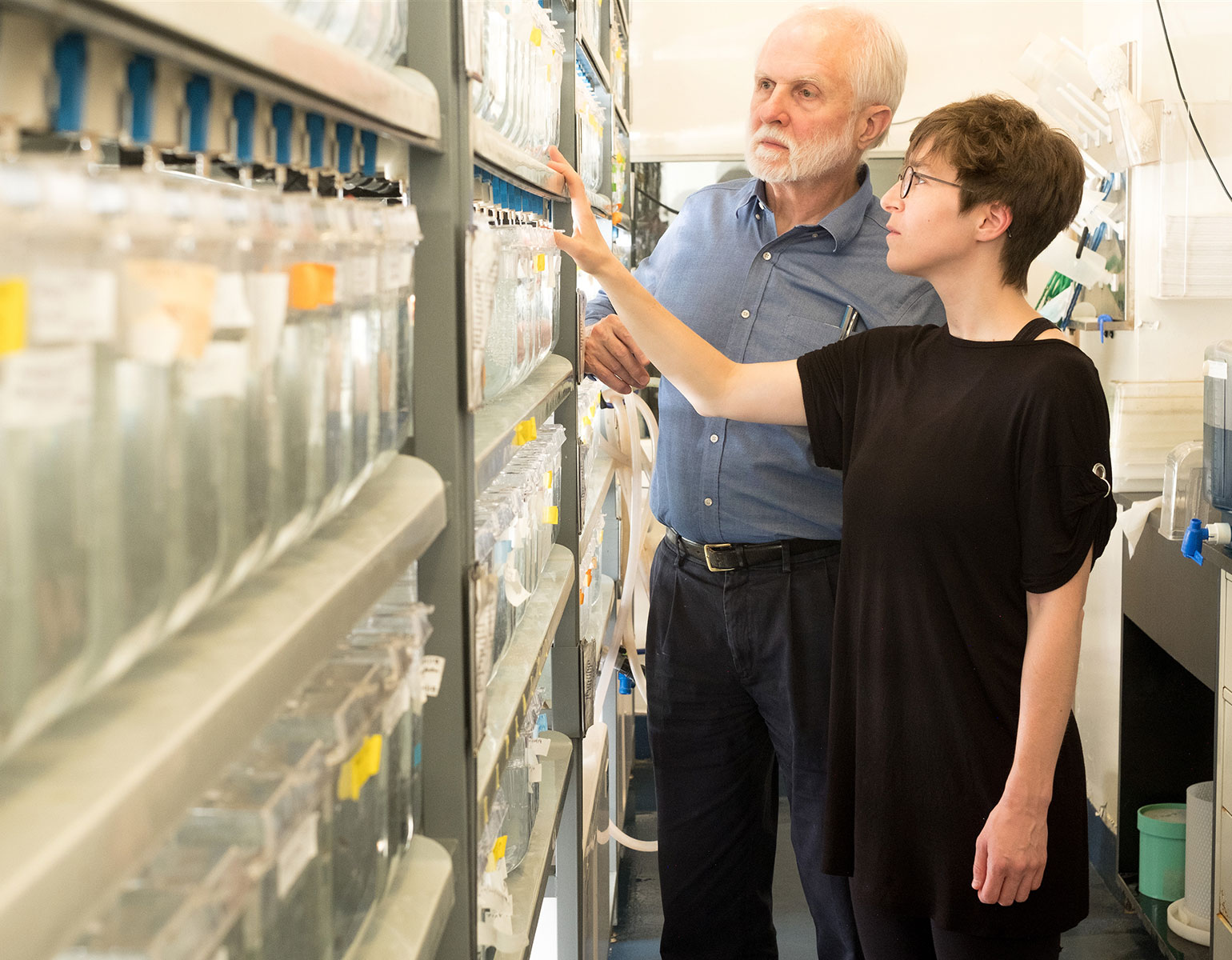
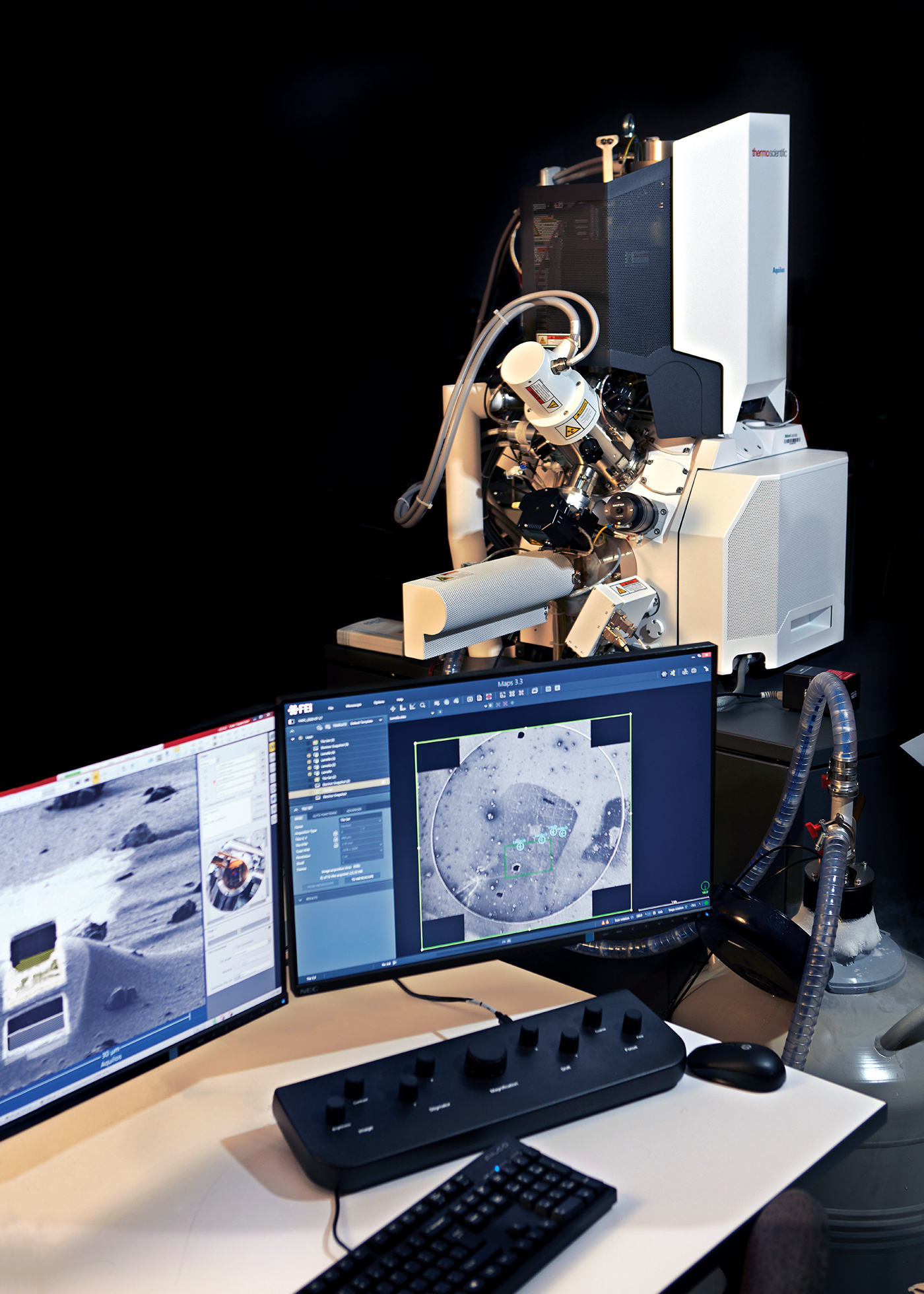
An intriguing example of such cellular zombies was recently discovered in the lab of Paul Greengard, a Nobel laureate and Rockefeller’s Vincent Astor Professor, who passed away last year. A team of scientists was trying to figure out what goes wrong in the brains of people with Parkinson’s disease. The researchers had long struggled to understand why dopamine-producing neurons in the midbrain perish, leading to debilitating movement problems characteristic of Parkinson’s.

When zombies take over the brain

Data

Neuro­degener­ation happens in all people, all the time. On average, an adult loses 3,250 neurons every hour.

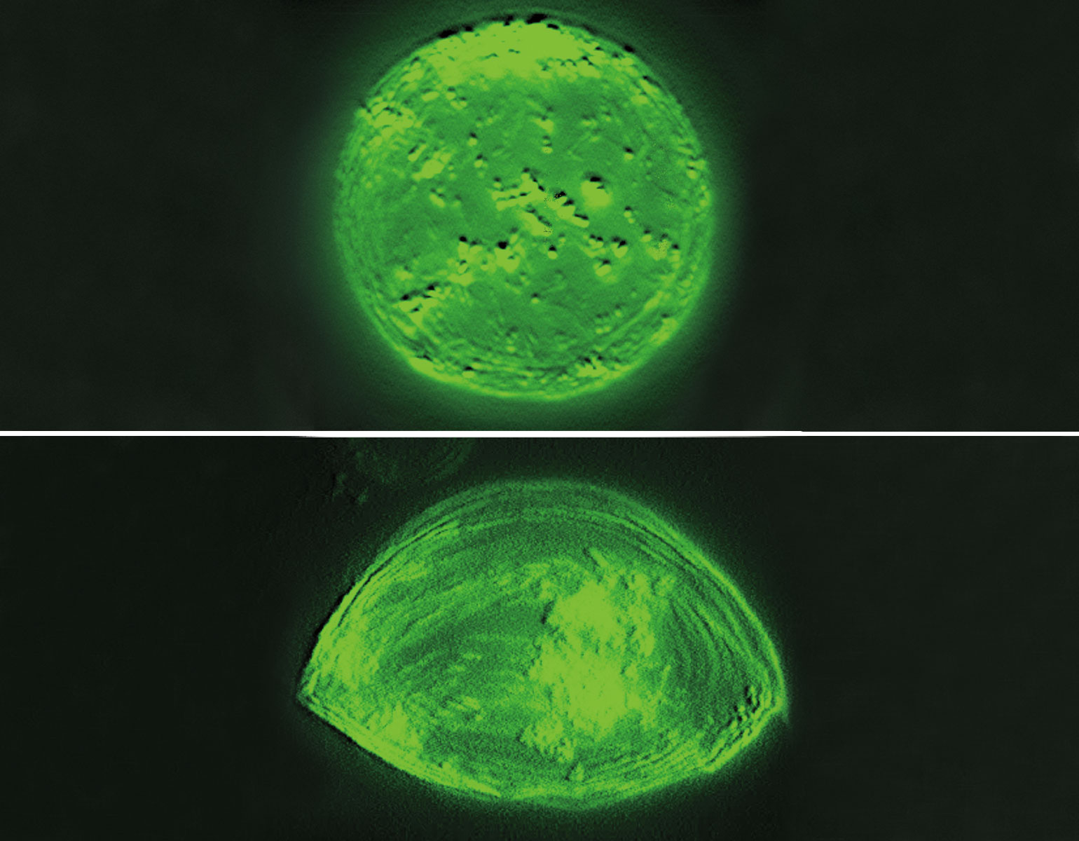
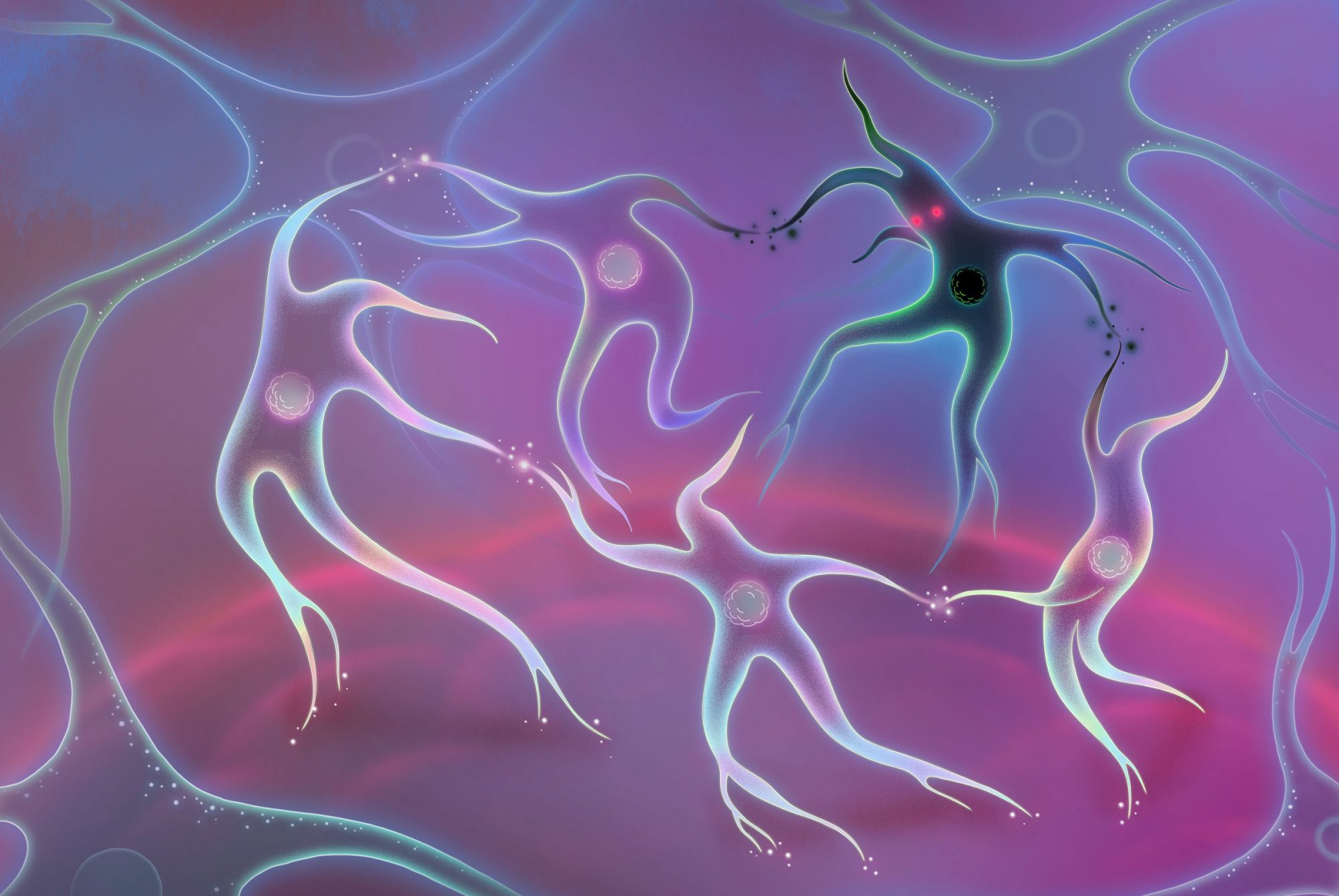
In retrospect, they may have been asking the wrong question. As the scientists reported in Cell Stem Cell, at least some of these midbrain neurons appear not to be dead after all, but rather to be resting in a zombie-like state known as senescence. And the results suggest that a zombie neuron may be even more damaging to the central nervous system than a dead one: By releasing inflammatory chemicals, the undead cells spread senescence to surrounding healthy neurons and make those neighbors shut down as well.

Research associate Markus Riessland says the discovery was especially surprising given that senescence is almost unheard of among neurons, although it does occur frequently in other parts of the body.

“Our findings shed new light on how Parkinson’s disease progresses,” he says, “and might provide new opportunities for treatment.” For example, Riessland and his colleagues suspect that so-called senolytic drugs, which are known to remove senescent cells, might make it possible to slow the brain’s deterioration.

Top-tier biosafety
Photograph by Frank Veronsky

On Campus

Top-tier biosafety

Photograph by Frank Veronsky

This lab is Hotel California for the tuberculosis bacterium, West Nile virus, SARS-CoV-2, and other deadly pathogensthey can never leave. For scientists, it’s the only place to study these highly infectious agents without infecting themselves or others. The 5,600 square-foot suite, known as a biosafety level 3 facility (BSL-3), uses high-volume HEPA filters and negative pressure to contain airborne pathogens. Constructed last year, it is one of only a handful of BSL-3 labs in New York City, and has proved to be a crucial resource for Rockefeller scientists studying the novel coronavirus (read more about their work here).

Some cells are multilingual
Erzberger and Hudspeth with tanks of zebrafish. Photograph by Mario Morgado

Forefront

Some cells are multilingual

Erzberger and Hudspeth with tanks of zebrafish. Photograph by Mario Morgado

When embryonic structures take shape, their cells must navigate with painstaking choreography. One wrong step, and entire sheets of tissue may warp. The hair cells of the inner ear, the sensory organ used for hearing and balance, are a striking example of such precision: They neatly line up in two rows facing each other, like cadets preparing for a drill.

“The cells have no blueprint for where to go, they just figure it out themselves by talking to each other,” says postdoctoral fellow Anna Erzberger. It sounds simple, but it’s not.

Together with colleagues in the group of A. James Hudspeth, the F. M. Kirby Professor, Erzberger has studied the developmental process that plays out in the fish equivalent of an inner ear. As it turns out, immature hair cells use more than one language to communicate.

Fish use a vibration-sensing organ similar to the ear to detect predators’ movements in the water around them.

When one cell divides into two, the daughters first engage in biochemical signaling—long held to be the singular mode of cellular discourse—to establish their individual identities. But soon after, they switch to mechanical lingo, enabling a kind of course correction. At this stage, the two cells may randomly find themselves occupying either the “right” or the “wrong” spot relative to one another—and in the latter case, they swap places. Propelled by surface-tension forces, the two cells gingerly dance past each other to assume the correct orientation.

“Traditionally, scientists have looked only to changes in genes and proteins to explain how developmental events happen,” says Erzberger, who coauthored a paper on the findings published in Nature Physics in May. “But biochemistry is only part of the story, and the missing link is often mechanics.”

A ride out of the pandemic?
For years, Rocky and Marley have participated in research aimed at harnessing the wonders of their immune system. The llamas live in pastures in rural Massachusetts. Photograph by Capralogics

Forefront

A ride out of the pandemic?

For years, Rocky and Marley have participated in research aimed at harnessing the wonders of their immune system. The llamas live in pastures in rural Massachusetts. Photograph by Capralogics

“It seems very strange that we should be picking, of all things, llamas,” says Michael P. Rout, referring to his latest project with Brian T. Chait, the Camille and Henry Dreyfus Professor. “But for reasons we don’t really understand, llamas make antibodies with fantastic properties.”

Llama antibodies are smaller than those of humans make but just as potent, and also easy and cheap to produce. The two scientists—with help from their two llamas, Rocky and Marley, are exploring antibody-based COVID-19 treatments with advantages that similar drugs based on human antibodies don’t have, such as the potential to scale up globally.

Watch this video to learn more.

We need to get better at making vaccines, and not just because of COVID
Activated B cells cluster in germinal centers (blue, green, and purple) inside a mouse lymph node. The Rockefeller University / Laboratory of Lymphocyte Dynamics

Forefront

We need to get better at making vaccines, and not just because of COVID

Activated B cells cluster in germinal centers (blue, green, and purple) inside a mouse lymph node. The Rockefeller University / Laboratory of Lymphocyte Dynamics

New pandemics require new vaccines, but so do old plagues. For example, scientists have tried for decades to develop a universal flu vaccine that works for every version of the virus, but this goal remains elusive. Last year, influenza killed more than 60,000 Americans.

But new opportunities are on the horizon—and according to Gabriel D. Victora, the Laurie and Peter Grauer Assistant Professor, some might come from learning how antibody-producing B cells move in and out of germinal centers. Located in lymph nodes, germinal centers are what Victora calls “boot camps” for activated B cells. “The cells go in as amateurs and come out as skilled professionals making antibodies that bind more tightly to their targets,” he says.

In recent work published in Cell, Victora’s team found that those educated B cells don’t easily return to camp the second time they’re exposed to an invader—which might make it hard to develop effective vaccines against highly variable viruses. “If we can find the bottlenecks, and learn how to circumvent them, that might lead to improved vaccination strategies,” he says.

The implications may extend to viruses other than the flu, such as HIV and hepatitis C—and perhaps also to coronaviruses.

This fish is about to flip
Science Photo Library

Forefront

This fish is about to flip

Science Photo Library

It’s not pleasant to swim when the pool is too hot.

In the lab of Alipasha Vaziri, a gang of zebrafish larvae has found an elegant solution to this problem: By simply flipping their tails to one side, they can cool down the water around them. The direction of the flip is important—every once in a while, the fish will try flipping the other way. It never helps.

This fish is about to flip

Data

The number of neurons in an adult zebrafish. Cats have about 25 times as many; lobsters have 100 times fewer.

Outside the fish tank is a team of researchers who, unbeknownst to the fish, are guilty of warming the water in the first place, with lasers, and of letting it cool to reward fishes that have learned the “correct” way to flip. In repeating this drill, they’ve been teaching the fish a new, goal-oriented behavior. And once the animals have been trained, they become part of an intricate set of experiments designed to shed light on one of neuroscience’s greatest enigmas: how brains make decisions.

When a fish responds to rising temperature, it flips in the correct direction about eight times out of ten. The researchers closely monitor each tail flip while simultaneously detecting the activity of neurons in the animals’ brains. The whole episode takes about 20 seconds, but the scientists are homing in on a shorter interval right after the water-warming laser is switched on and before the fish moves—the key moment when the left-or-right choice is made.

“The goal is to understand how decisions unfold,” says Vaziri.

In their findings, recently published in Cell, the scientists describe the activity state of about 5,000 neurons across the entire fish brain. They identified a number of activity patterns—some related to the brain’s sensing the heat, some to its coordinating tail flips, and others to the decision-making process. They also found that about 10 seconds before a fish moves, those patterns will foreshadow whether it’s about to make the correct or incorrect turn.

How brains make decisions is one of neuroscience’s greatest enigmas.

In fact, just by observing the brain-activity profiles, the scientists could usually guess beforehand when the fish would move its tail, and whether it would gear left or right.

If predicting an animal’s next move seems remarkable, so is the technology the scientists built in order to conduct these experiments. Tracking how neurons across multiple brain regions respond and cooperate is anything but trivial, and Vaziri’s team made it possible by pairing advanced statistical methods with a novel light-field microscopy technique developed in the lab.

A mediocre mutator
SARS-CoV-2 particles isolated from a COVID-19 patient are docking onto a host cell (lower right). The virus uses its spike proteins (orange) to attach to the cell and break into it. National Infection Service / Science Photo Library

Forefront

A mediocre mutator

SARS-CoV-2 particles isolated from a COVID-19 patient are docking onto a host cell (lower right). The virus uses its spike proteins (orange) to attach to the cell and break into it. National Infection Service / Science Photo Library

It’s a nightmare pandemic scenario: The drugs and vaccines being developed for COVID-19 are rendered useless by a SARS-CoV-2 mutation, and the scientists working on these treatments go back to square one. What if, like HIV or influenza, the coronavirus will be able to tweak its genome to dodge medical defenses and stay a step ahead of science? The possibility has been keeping people awake at night ever since the virus first escaped Wuhan.

But new research has delivered hopeful results, suggesting that SARS-CoV-2 may not be the master escape artist those other viruses are. A team of Rockefeller scientists found that, although the virus could potentially accumulate mutations affecting its spike protein—the key viral molecule recognized by antibodies—there are ways to prevent such mutants from resisting future treatments.

Scientists in the labs of Paul BieniaszMichel C. Nussenzweig, and Charles M. Rice are developing therapies for COVID-19 based on antibodies harvested from patients who successfully overcame the coronavirus. They are banking that these antibodies—amplified in the lab using cloning techniques—will bind to the spikes, preventing the virus from entering human cells (read more about their work in “Inside the Response”).

A mediocre mutator

Data

A single nasal swab from a person infected with SARS-CoV-2 can contain 1 billion copies of viral RNA.

Whether such drugs will remain effective over time depends on the likelihood that mutations will change the sequence and structure of the viral spike. So the team designed a series of experiments to see whether the spike could acquire resistance to the therapeutic antibodies. In findings published in August on the preprint server BioRxiv, they combined a faux coronavirus expressing the SARS-CoV-2 spike protein with antibodies, and grew the virus in human cells in a dish, then observed changes in the spike protein.

Of a vast pool of potential viral mutants, a small fraction was selected that dodged the antibodies and was able to infect cells. Predictably, these escapees carried slight genetic modifications in the spike protein, “the very types of mutations that could potentially make the virus resistant to antibody treatment,” says Theodora Hatziioannou, a research associate professor in the Bieniasz lab.

But although some resistant mutants arose in the presence of individual antibodies, none were detected when a cocktail of two different antibodies targeting distinct spike regions was used. That is reassuring, Hatziioannou says, as it suggests that a drug formulation combining two or more antibodies would be unlikely to fail.

The scientists are also working to determine the probability that spike-protein mutations will undermine the effectiveness of a future vaccine. Quantifying that risk is more complicated, Hatziioannou says, since vaccines are typically designed to make the body produce its own antibodies, as opposed to introducing a specific kind of antibody into a patient’s bloodstream. Success would therefore depend on what type of antibody response a given vaccine candidate elicits and how that response varies among people.

Further clues will begin to emerge as data from large-scale phase III clinical trials of developmental vaccines, now underway internationally, become available.

Attention needs more attention
Illustration by Chris Kindred

Forefront

Attention needs more attention

Illustration by Chris Kindred

Life can sometimes feel overwhelming—and it literally is. Every second, a tremendous soup of sensory information floods the human brain: sights, sounds, and sensations, all clamoring to be known. If not for the brain’s capacity for selective attention, the world would forever look like chaos.

Selective attention allows the brain to decide which sensory input to prioritize at any given moment (as you’re reading these words, for example, it’s giving premium processing status to the sight of letters). It’s an essential task whose biological machinery resides in a handful of areas, all confined to the brain’s parietal and frontal lobes—or so scientists have long thought.

A few months ago, however, two neuroscientists reported their discovery of a new area that appears to control selective attention. In their behavioral experiments, in which subjects were tasked with watching moving dots on a screen, this area kept activating. Further tests revealed that neurons in this area closely track precisely which part of the screen is being attended to—the signature characteristic of an attention-governing brain region.

The discovery has introduced new mysteries. For one thing, the new area is located in an unlikely place—the dorsal part of the posterior inferotemporal cortex—that has not previously been linked to attention. It suggests that the classical account of selective attention isn’t the full story, says Rockefeller professor Winrich Freiwald, who published the findings in the Proceedings of the National Academy of Sciences together with a colleague at the University of Bremen.

“Scientists may have to fundamentally rethink how some aspects of our brains are organized,” Freiwald says.

Primordial shapeshifters
Synthetic cells have mastered the art of stretching, a prerequisite for self-replication.

Forefront

Primordial shapeshifters

Synthetic cells have mastered the art of stretching, a prerequisite for self-replication.

It’s the holy grail of synthetic biologists: creating a living cell from scratch. So far they’ve managed to make simple prototypes—essentially tiny fat balloons with a soup of genetic material inside, capable of reading genetic code, producing proteins, and transporting molecules around. Yet these artificial blobs lack an essential feature shared by all living things: the ability to generate more copies of themselves.

Self-replication is arguably the most sophisticated of biological phenomena and has long seemed nearly impossible to engineer. But clues are starting to emerge thanks in part to Albert J. Libchaber, the Detlev W. Bronk Professor Emeritus, who became interested in the process by which a cell deforms from a sphere into an oval—a first step required for it to split into two. “It’s not easy to divide a perfect sphere,” he says.

Together with Vincent Noireaux, a postdoc in the lab now at the University of Minnesota, Libchaber found a secret ingredient that can help cell prototypes elongate: polyethylene glycol, a sticky molecule found in skin creams and soap bubbles. Previously, the scientists had tried stretching their spherical creations with MreB, a protein that builds a bacterium’s inner scaffolding, which molds the cell into its trademark rod-like shape. But MreB on its own did nothing to flatten Libchaber’s cell replicas; only after polyethylene glycol was added did it turn into a dynamic polymer capable of inducing the sphere-to-oval transformation.

The findings, published in the Proceedings of the National Academy of Sciences, bring scientists a step closer to creating a self-reproducing molecular contraption: a truly living cell made from 100 percent dead ingredients.

What Darwin never guessed
Turtle ant soldiers display a wide range of head shapes and sizes. Photograph by Scott Powell

Forefront

What Darwin never guessed

Turtle ant soldiers display a wide range of head shapes and sizes. Photograph by Scott Powell

The origin of species usually goes like this: One group of stickleback fish lives at sea, the other goes to freshwater, and voila—one species becomes two. For centuries, evolution has been thought to generally march in the same direction, toward new traits and more-specialized adaptations. But recently, scientists at Rockefeller and George Washington University found a quirky exception.

Turtle ants live in trees, in tunnels previously excavated by beetles. To keep intruders out, ant soldiers use their large heads to plug the tunnels. But when an ant colony moves to a new habitat, the soldiers may have to adapt to larger or smaller holes dug by a different beetle species, and the size and shape of their heads evolves for a snug fit. Surprisingly, researchers found that this aspect of ant evolution has gone both forward and backward—sometimes creating novel head shapes, other times reverting to more primitive ones.

“You would think that once a species is specialized, it’s stuck in that narrow niche,” says Daniel Kronauer, the Stanley S. and Sydney R. Shuman Associate Professor, whose team published the findings in the Proceedings of the National Academy of Sciences. “But turtle ants have a very dynamic evolutionary trajectory with a lot of back and forth.”

Benign buds
The Rockefeller University / Robin Chemers Neustein Laboratory of Mammalian Cell Biology and Development

Snapshot

Benign buds

The Rockefeller University / Robin Chemers Neustein Laboratory of Mammalian Cell Biology and Development

Two of the most common forms of skin cancer arise from the same sourceepidermal stem cellsbut take different paths in life. Basal cell carcinomas start off as bud-shaped cell clusters and tend to humbly stay put. This makes them less aggressive than their cancerous cousins, squamous cell carcinomas, which originate as tiny folds before burrowing into deeper layers of the skin to form tumors capable of spreading throughout the body.

A team led by Elaine Fuchs, the Rebecca C. Lancefield Professor, captured this image while trying to understand what makes some precancerous tumors turn malignant while others remain relatively benign. It turns out that the mechanical properties in the tissue are key factors determining whether epidermal stem cells (green) grow up to become the docile buds seen aboveor insidious folds in the skin, just waiting to metastasize.

Return of the cytonaut
Illustration by Wenkai Mao

Forefront

Return of the cytonaut

Illustration by Wenkai Mao

“Cells live in a watery world, even when the organisms of which they are part do not,” wrote Christian de Duve in his 1984 book, A Guided Tour of the Living Cell. Most human cells, for instance, are immersed in fluids that render their vast inner space jelly-like. Floating within this cytoplasmic soup are mitochondria, ribosomes, vacuoles, and many other so-called organelles—tiny instruments that carry out a cell’s basic functions.

Inspired by the films of Jacques Cousteau, de Duve wanted his readers to embark on the journey with the mindset of a “cytonaut” ready to explore “a strange world, fascinating, mysterious, but very far removed from our everyday experience.” The late cell biologist was himself a pioneer who, together with Albert Claude and George E. Palade spent the 1940s and 1950s detailing the first functional map of the cell—work for which the three Rockefeller scientists later shared a Nobel Prize.

A must-see along the tour is the lysosome, a bubble-shaped organelle that de Duve was the first to set eyes on in 1955, and whose acidic interior was subsequently found to serve various purposes, such as breaking down cellular debris. Yet it wasn’t until early this year that scientists discovered that our cells need this sour little sac to process iron, an essential nutrient, into a form they can metabolize in order to survive. This may, in fact, be the most important of the lysosome’s functions.

Graduate student Ross Weber made the discovery in a lab not far from the one de Duve once inhabited. There, in an experiment that had to be controlled with minute precision, he manipulated cells to make their lysosomes less acidic. For reasons that have long been unknown, cells will stop dividing and die if the pH within lysosomes rises above a certain threshold.

Today Weber and other members of the lab, led by Kivanç Birsoy, have a possible explanation for this phenomenon. Their experiments show that cells with more-alkaline lysosomes suffer iron depletion—and as a result, they lose their ability to produce essential molecules such as DNA. “Lysosomes participate in a lot of different cell functions like signaling, metabolism, and recycling,” says Birsoy, who is Rockefeller’s Chapman Perelman Assistant Professor, “but processing iron seems to be the only thing cells really cannot do without them.”

He hopes the new research, published in Molecular Cell, might lead to the development of novel cancer therapies. Several types of tumor cells are known to be sensitive to elevated lysosome pH, and the new findings suggest it’s the ensuing iron deficiency that deals these tumor cells a fatal blow. This could mean that depleting tumors of iron offers an effective way to kill them, says Birsoy. This is the latest possibility to come out of his lab’s extensive effort to develop new treatments that starve tumors for nutrients they cannot produce on their own.

The team also plans to explore whether the new findings could be relevant to other conditions linked to the loss of lysosome acidity, including a group of rare metabolic disorders and neurodegenerative diseases. “We believe there are a lot of exciting possibilities out there,” Birsoy says.

Moreover, the lysosome isn’t the only organelle whose inner secrets might yield ideas for new medicines. Mitochondria, for example, the cell’s peanut-shaped powerhouses, are the targets of several promising cancer treatments. And who knows what other treasures await 21st century cytonauts as they plunge deeper into the cellular sea.

Scope and scalpel in one
Photograph by Matthew Septimus

Gadget

Scope and scalpel in one

Cryo-electron microscopy can make the invisible visible, but you have to know where to look. It’s a bit like pointing your telescope into the sky; there’s a lot of darkness out there in between the interesting parts.

The cryo-electron focused ion beam milling microscopethe cryo-FIB for shortis like a star finder. Although it lacks the power of a full-fledged cryo-electron microscope, which uses extremely cold temperatures to “fix” samples for imaging, the cryo-FIB has a wider field of view and comes with sophisticated tools for manipulating samples to get the best view. Scientists can use it to identify areas of interest and precisely orient them for study in larger machines. It makes hours spent in the cryo-EM rooms both more efficient and more productive.

The best feature: the cryo-FIB’s focused ion beam, which can slice off razor-thin sheets of atoms with nanometer precision, uncovering new molecules of interest.

“It’s like a deli slicer,” says Mark Ebrahim, senior staff scientist in the Evelyn Gruss Lipper Cryo-Electron Microscopy Resource Center. “It trims the cell layer by layer until you get the specific slice you need.”

For structural biologists, who use cryogenic technologies to understand how a cell’s tiniest components function and to design novel drugs, the possibilities are now seemingly endless.