Genetic diversity comes into focus
Illustration by Pierre Buttin

Forefront

Genetic diversity comes into focus

Illustration by Pierre Buttin

The humble wild cabbage has had one since 2016. A lowly gut bacterium has had one even longer. Now, courtesy of the Human Pangenome Reference Consortium—an international alliance whose leaders include Rockefeller’s Erich D. Jarvis—we Homo sapiens finally have one too: a genetically diverse pangenome that promises to dramatically increase our understanding of human disease and expand access to personalized medicine.

When the human genome was first released in 2003, scientists were already working to improve it. Over the next two decades, technology advances made it possible to fill in gaps and correct errors, but a substantial problem remained: Two-thirds of the DNA in the original reference genome came from a single person. As a result, many genetic variants found in non-European populations, such as people of African or Asian descent, weren’t included.
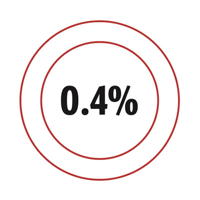
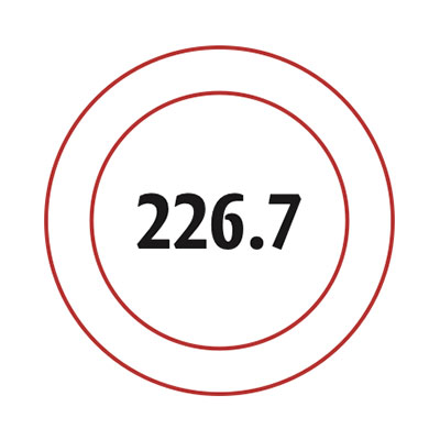
.4 percent

Data

The fraction of human DNA that varies from person to person.

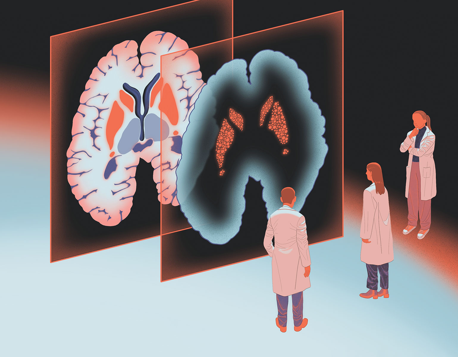
This lack of representation can lead to biases in biomedical data that may in turn contribute to inequities and health disparities between different groups, Jarvis says. Among people with European ancestry, researchers have discovered countless genetic variants that predispose to specific illnesses, influence the severity of disease, or affect responses to particular drugs—knowledge that can provide powerful tools for physicians to diagnose diseases, predict health outcomes, and tailor treatments for individual patients. Such discoveries have yet to be made for populations whose variants were excluded from the reference genome in the first place.

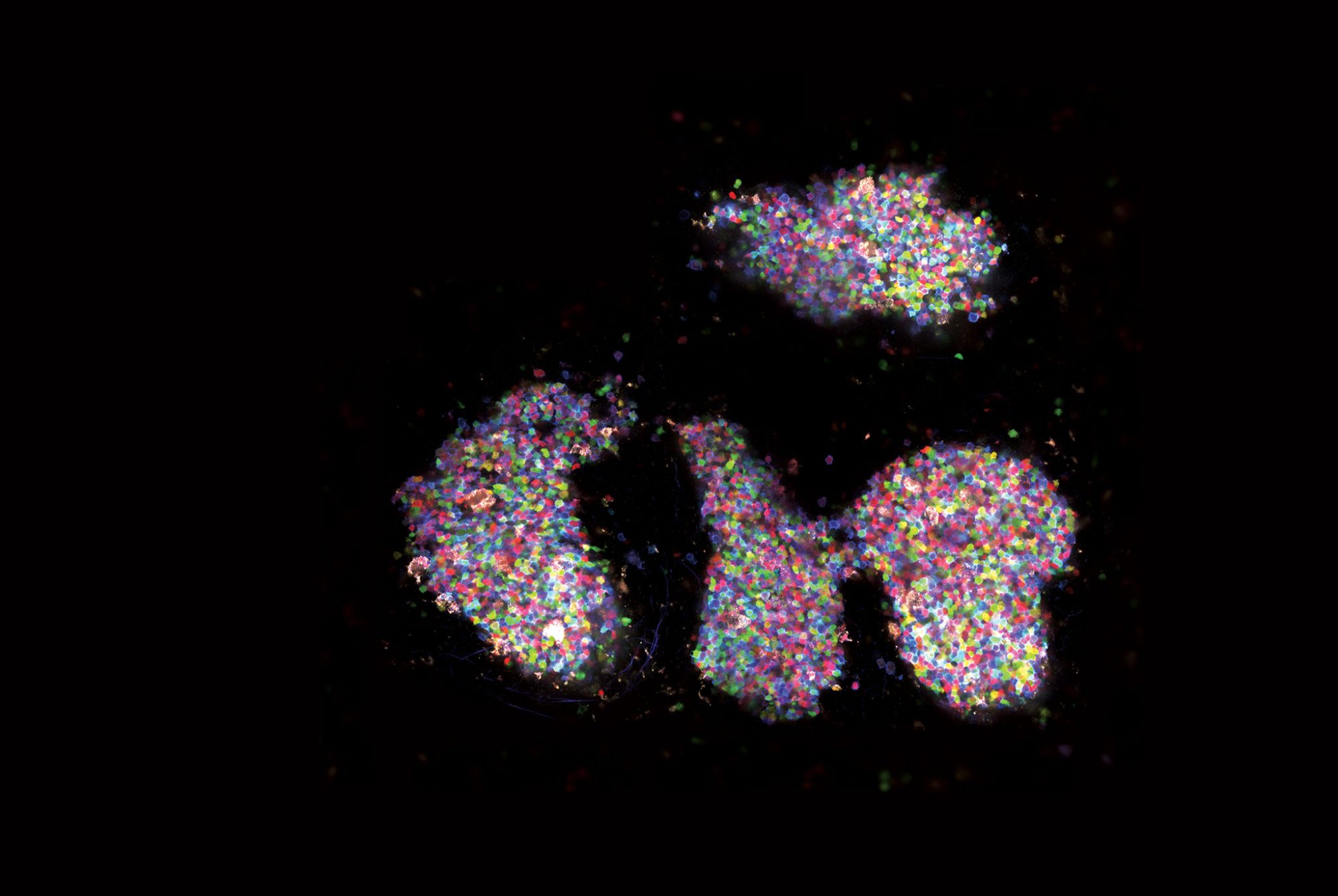
The human pangenome that Jarvis and his colleagues have unveiled unveiled is a first step toward tackling these problems head-on. Obtained with DNA from 47 people from around the world, it reveals nearly 120 million new DNA data points. Most are related to so-called structural variations, genetic differences that arise when long stretches of the double helix are duplicated, deleted, or rearranged.

“Structural variations can have dramatic effects on trait differences, disease, and gene function,” Jarvis says. “There will be a lot of new discoveries to come that weren’t possible in the last 20 years.”

A garden where kids can grow
Photo by Matthew Septimus

On Campus

A garden where kids can grow

Photo by Matthew Septimus

There are few spaces in this dense city where children can make mud pies and pluck raspberries, let alone learn STEM basics by planting and tending flora. But a campus garden has been a periodic Rockefeller feature since 1911, at one point swelling to two acres. Nowadays, 102 toddlers in the university’s childcare and preschool enjoy a smaller (albeit large for Manhattan) 706-square-foot plot. Lovingly tended by teachers, parents, and the kids themselves, the harvest is used in countless classroom science projects—when it isn’t eaten right off the vine.

Brain cell biographies

Forefront

Brain cell biographies

Our brains continually produce new cells, a generative process that slows down as we reach our golden years. With new technology, scientists are now able to capture the ebb and flow of various brain cell types during normal development and aging and investigate what happens to decaying cells in neurodegenerative diseases like Alzheimer’s.

Geneticist Junyue Cao and his colleagues zeroed in on progenitor cells, descendants of adult stem cells that differentiate into specialized cell types. By attaching unique ID tags to more than 10,000 newborn progenitor cells in the brains of mice, they were able to track these traditionally elusive cells and study their fates throughout the animals’ life span. “It’s like an ID card and GPS tracker combined,” Cao says of the new technique, called TrackerSci, which offers wide-ranging applications. “If we can systematically characterize the different cells and their dynamics, we may get a panoramic view of the mechanisms of many diseases and the enigma of aging.”

Tiny lung buds for big experiments
LABORATORY OF SYNTHETIC EMBRYOLOGY

Forefront

Tiny lung buds for big experiments

LABORATORY OF SYNTHETIC EMBRYOLOGY

It’s been hard to figure out how SARS-CoV-2 wreaks havoc in the lungs, partly because many studies have been done in samples from patients who respond differently to the virus. If you’re trying to pinpoint a disease mechanism, you want to run many comparable trials side by side, not deal with a hodgepodge of genetically diverse situations.

That’s why the labs of Ali H. Brivanlou, the Robert and Harriet Heilbrunn Professor, and Charles M. Rice, the Maurice R. and Corinne P. Greenberg Professor, developed a cell-culture platform capable of growing and infecting millions of lung buds, minuscule structures resembling those that give rise to our breathing organs. With this technology, they hope to light up COVID’s attack route like an airport runway. In experiments described in Stem Cell Reports, for example, the researchers found that alveoli, the tiny sacs at the end of lung branches, are more susceptible to infection than cells in the airway, which are the first line of defense against inhaled pathogens. If the virus gets past the airway, the alveoli are sitting ducks.

“This technology is ready to confront all kinds of threats,” Brivanlou says, from respiratory infections to noninfectious diseases like lung cancer. “It can be used to screen drugs, vaccines, monoclonal antibodies, and more, directly in human tissue.”

His creative thinking transformed our understanding of gene regulation

Forefront

His creative thinking transformed our understanding of gene regulation

C. David Allis was never afraid to buck conventional wisdom.

In the 1980s, most scientists thought that histones, the proteins around which DNA is wrapped, were little more than packaging material for the molecule of life. Together, histones and DNA form a substance called chromatin, and researchers believed the histone part was basically the bubble wrap. Allis enthusiastically threw himself into studying these seemingly uninteresting proteins using Tetrahymena, an obscure single-celled organism. Many of his colleagues thought he was wasting his time.

“But of course, what he discovered in that little organism would turn out to be relevant to all of us,” says Robert G. Roeder, the Arnold and Mabel Beckman Professor.

Allis found that histones play a critical role in turning genes on and off and in fine-tuning their effects—a breakthrough that revolutionized our understanding of how the basic instructions encoded in DNA are expressed in our tissues. Moreover, his research offered fresh insight into diseases as disparate as cancer and dementia and paved the way for new treatments.

He was also known for his humor, gentle demeanor, and dedication to his students and postdocs. “For someone so accomplished, he was the kindest, most humble, and relentlessly positive person you could imagine,” says Richard P. Lifton, the Carson Family Professor and president of The Rockefeller University.

Allis, the Joy and Jack Fishman Professor, died in January last year. He was 71.

Where credit is overdue
Photograph by Chris Taggart

Forefront

Where credit is overdue

Photograph by Chris Taggart

Ozempic, Wegovy, Saxenda, Victoza—these blockbuster drugs for treating diabetes and obesity have all but become household names. But what about the name Svetlana Mojsov?

A research associate professor at Rockefeller, Mojsov laid the scientific groundwork that made these treatments possible (learn more about her work in “A drug’s discovery”). While working at Massachusetts General Hospital in the 1980s, she discovered glucagon-like peptide 1, or GLP-1, a hormone secreted by the gut that triggers insulin production and lowers blood sugar. The new drugs—which began as diabetes treatments and were later found to induce weight loss as well—mimic the hormone’s effects. The first to come on the market, Victoza for type 2 diabetes and Saxenda for weight loss, were based on the GLP-1 sequence that Mojsov discovered.

3 to 12 percent

Data

The typical fraction of total body weight lost after a year of treatment with GLP-1-based drugs.

Her work was a godsend to drug developers. Safe, effective weight-loss treatments have long eluded researchers. Many drugs have had to be pulled from the market after causing life-threatening side effects or hard-to-kick addictions. The new class of GLP-1 agonists operate in a fundamentally different way, however, and are now being reliably used by millions across the world to lose weight and manage diabetes.

But while other researchers have received major awards for contributing to the development of these drugs, Mojsov remained unrecognized. Over the years, she has had to fight to have her name included on GLP-1 patents as a coinventor, and correct papers in high-profile journals that didn’t acknowledge her work.

Now, that long overdue credit is finally rolling in. And while Mojsov is glad that she is no longer faced with the prospect of being erased from scientific history, what matters most to her is that these drugs are helping to improve the health and well-being of millions of people. “That makes me feel professionally and personally fulfilled,” she says.

Hunger games for tumors
PHOTO BY CLAIRE HOLT

Forefront

Hunger games for tumors

PHOTO BY CLAIRE HOLT

Cancer scientists may have hit on a way of forcing tumors to self-sabotage. It emerged from experiments in which a team led by Sohail Tavazoie, the Leon Hess Professor, examined cancer cells that were running low on arginine, an amino acid present in protein-rich foods.

These malnourished cancer cells pursued several coping strategies during their lifespan, including accruing a DNA error that made their offspring less arginine-hungry. Experiments described in Science Advances showed that the longer the cells grew without arginine, the more these mutations piled up. And in theory, the more mutations a tumor has, the more likely it is to be detected and destroyed by the body’s immune cells.

Dennis Hsu, a former member of Tavazoie’s Elizabeth and Vincent Meyer Laboratory of Systems Cancer Biology who is a physician-scientist at UPMC Hillman Cancer Center in Pittsburgh, suspects that cancer cells’ dependence on arginine could thus be leveraged to make tumors more vulnerable to immunotherapy drugs that rally the immune system to destroy weird-looking cells. Withdrawing arginine might make it possible to trigger a rash of tumor mutations, essentially painting an immunological bull’s-eye on the cancer cells and obviating the need to attack them with toxic chemicals or radiation.

“We haven’t tested this yet,” Hsu says, “but it would be a really cool thing to try.”

Smelling danger
A transgenic ant pupa flashes neon in the presence of alarm pheromones. LABORATORY OF SOCIAL EVOLUTION AND BEHAVIOR

Forefront

Smelling danger

A transgenic ant pupa flashes neon in the presence of alarm pheromones. LABORATORY OF SOCIAL EVOLUTION AND BEHAVIOR

Whether foraging, fighting, or parenting, ants are continuously sending and receiving smell signals called pheromones. Recently, scientists used a glowing protein derived from a bioluminescent jellyfish to ask which parts of an ant’s brain responds when it catches a whiff of an alarming scent.

Daniel Kronauer, the Stanley S. and Sydney R. Shuman Associate Professor, and his team injected the eggs of clonal raider ants with genetic material encoding the protein GCaMP, which glows neon green when calcium levels change due to cellular activity. They aimed these proteins at specialized brain structures called glomeruli, which are essential to scent processing. They then used a custom imaging technique to monitor GCaMP levels in the glomeruli as the ants took in a range of odors. Alarm pheromones caused six glomeruli in one particular region to light up, suggesting that this area may act as the brain’s panic button.

The technique, described in Cell, could potentially be used to reveal what hundreds of odorant receptors are up to, says first author Taylor Hart, a postdoctoral associate in Kronauer’s lab who has bred hundreds of such gleaming ants.

“This opens up a big range of questions that were inaccessible to us until now,” adds Kronauer.

Decision time
Egg-laying neurons (in green) light up as a fruit fly makes a decision. JANELIA FLYLIGHT TEAM

Forefront

Decision time

Egg-laying neurons (in green) light up as a fruit fly makes a decision. JANELIA FLYLIGHT TEAM

If a rock comes whizzing at your face, you’ll duck without a thought. But if you’re scanning the menu at a well-rated restaurant, you might spend several minutes weighing your gustatory options.

Both represent decisions, but while one occurs over a few seconds or less, the other takes much longer. And while neuroscientists have learned a lot about the mechanisms governing reflexive decisions, they know far less about those that operate over longer timescales.

Recently, Gaby Maimon’s Laboratory of Integrative Brain Function gained insight into how these slower decision-making processes unfold in the fruit fly brain as female flies choose a good spot to lay their eggs. In work published in Nature, the researchers homed in on a set of cells known as oviposition descending neurons, which play a key role in the process.

222 point 7

Data

Estimated number of food-related decisions a person makes in a given day.

They identified a calcium signal in these neurons that fluctuated as flies inspected different egg-laying options. The signal peaked at exactly the moment a fly began laying eggs, indicating it had crossed some kind of decision-making threshold.

Choosing where to lay eggs is not a reflexive decision but a considered one. In experiments where female flies were placed on a rotatable treadmill and allowed to walk across different surfaces mimicking fruits they might encounter in the real world, the little critters often took up to a minute to choose just the right spot for their eggs.

Pregnant flies spent up to a minute pondering where to lay their eggs.

And when first author Vikram Vijayan, a research associate in Maimon’s lab, inhibited the insects’ oviposition descending neurons, prolonging the time it took for the calcium signal to reach threshold, the flies took even longer to decide on a spot. The extra time benefited them: They laid more eggs on the surface that matched their expected preference in the wild.

“The more time the flies spent exploring,” Vijayan says, “the more they tended to pick an option that presumably ensured better survival of their offspring.”

The team’s findings could build a foundation for understanding how humans make educated and strategic decisions, says Maimon: “This work allows us to imagine that a similar rise-to-threshold process might exist in our own brain as we select what clothes to wear in the morning.”

Nightmare scenario

Forefront

Nightmare scenario

one day, during his late morning nap, Costello the octopus had a nightmare.

Video footage captured by cameras in biophysicist Marcelo O. Magnasco’s Laboratory of Integrative Neuroscience showed that while Costello began his snooze peacefully hanging from the side of his glass tank, the little octopus suddenly flushed with color and fell, thrashing and twisting, to the pebbled floor. He flexed his mantle into a cone shape, a defensive posture against predators; wrapped himself around a PVC pipe (one of his many toys) as if subduing a nemesis; and ended the fracas with a dramatic squirt of black ink. Eight minutes later, he was milky-hued and calm again.

The researchers were amazed when they reviewed the footage the next day. A Brazilian reef octopus, Costello had been captured off the coast of Florida with a missing arm—evidence of a battle he’d at least partially lost. Had he been dreaming about that altercation or a similar one? If so, what did that mean for octopus cognition, the focus of their research?

Just like human beings, octopuses have active sleep states during which they ignore external stimuli. But scientists don’t yet know whether they dream as we do, melding memory and invention into a full-blown narrative.

Costello had three more such episodes over the next month before dying of natural causes. Since publishing an article exploring the implications of this unusual behavior as a preprint on bioRxiv, Magnasco has continued to study octopus cognition (read more about his work in “The octopus examination room”).

Sleep occurs in virtually all animals, while dreaming has long been thought to be confined to neurologically complex vertebrates. But Costello’s episodes are an intriguing suggestion that our distant kin—and perhaps other spineless animals—may in fact be capable of complex sleep.

“If invertebrates dream,” Magnasco says, “then perhaps dreaming exists throughout the tree of life.”

An early flag for Parkinson’s
Illustration by Jun Cen

Forefront

An early flag for Parkinson’s

Illustration by Jun Cen

A woeful catch-22 plagues efforts to treat Parkinson’s disease: Early diagnosis can stave off the most severe symptoms for years; but without symptoms, doctors have no way of detecting the illness.

This is especially unfortunate because by the time symptoms do appear, sufferers have already lost 50–80 percent of the brain cells that produce dopamine, a neurotransmitter that plays a crucial role in various functions, from movement to memory.

Researchers are now looking for disease biomarkers that could be detected before symptoms surface. Thus far, their quest has been complicated by the challenges involved in examining brain tissue from living Parkinson’s patients.

So when Krithi Irmady, an instructor in clinical investigation, found overlapping RNA changes in the blood of living patients and the brains of deceased ones, she and her colleagues knew that they’d found something significant. The team’s findings, published in Nature Communications, could potentially be used to develop tools for predicting the course of the disease and creating treatment options tailored to a patient’s symptoms and disease stage.

The group discovered a bevy of RNA-driven gene-expression changes in one region of the brain associated with cognitive impairment and another region linked to motor control problems. Each bore distinct molecular signatures that could be linked to a patient’s symptoms—the first such markers to be found in Parkinson’s.

These and other recent findings by the team, which is headed by Robert B. Darnell, the Robert and Harriet Heilbrunn Professor, raise hope for the development of better drugs and prediction tools for the disease.

“I think our findings will generate excitement about the promise of blood-focused studies for Parkinson’s disease,” Irmady says.

When mutants mingle
LABORATORY OF LYMPHOCYTE DYNAMICS

Snapshot

When mutants mingle

LABORATORY OF LYMPHOCYTE DYNAMICS

One needs no scientific excuse to be entranced by a germinal center. This particularly lovely specimen belongs to a “Confetti” mouse whose cells were engineered to change color depending on how they rearrange their DNA, generating a spectacular display of the immune system at work.

Upon exposure to a pathogen, B cells cloister themselves inside germinal centers, which spring up inside lymph nodes, spleens, and tonsils, where the cells mutate over and over as they furiously refine their plan of attack. These elite fighters then emerge en masse, producing potent antibodies tailor-made to take out the infection.

The more we learn about germinal centers, the better we’ll understand how the body responds to disease and how to develop more-effective vaccines. To answer these questions, Gabriel D. Victora, the Laurie and Peter Grauer Associate Professor, is pinpointing the triggers that activate germinal centers and those that shut them down.

But gazing at this morass of color, we could almost forget that thousands of subtle, yet crucial biochemical reactions are taking place, driving immune cells toward perfection.

The octopus examination room
Photograph by Matthew Septimus

Gadget

The octopus examination room

Octopuses have secrets. In the wild, they’re homebodies who spend much of their time curled up inside a seashell, a coconut, or a rocky lair. In the lab, this poses a problem for neuroscientists like Marcelo O. Magnasco, who seeks a clearer picture of how octopuses perceive and interact with their environment.

These camouflaging, shape-shifting, sucker-spotted invertebrates are amazingly intelligent, with neural systems both markedly different from and strangely similar to ours, making them a unique model for studying how any brain engages with the world. Yet because an octopus’s behavior is largely hidden from view, the neural processes that drive it remain a mystery—a black box in our understanding of how the animal’s nine brains cooperate, and how its cognitive processes ultimately translate into sophisticated problem-solving skills and mischievous personalities.

Ironically, Magnasco’s solution is a literal black box, composed of the same black plastic once found in old-school TV remotes. Each of his lab’s six cephalopods has its own roomy tank filled with plants, stones, and toys. When a box is introduced, the octopus quickly adopts it as a cozy den. The cube is opaque to visible light but transparent to the infrared camera trained on it 24/7. This allows the inhabitant to feel unobserved while the researchers engage in “a gross invasion of privacy,” Magnasco jokes, enabling their exploration of the connection between learning and sleep—and even whether octopuses have nightmares. (Read more about cephalopod dreams in “Nightmare scenarios“).