Finding our human voice
Illustration by Harriet Lee-Merrion

Forefront

Finding our human voice

Illustration by Harriet Lee-Merrion

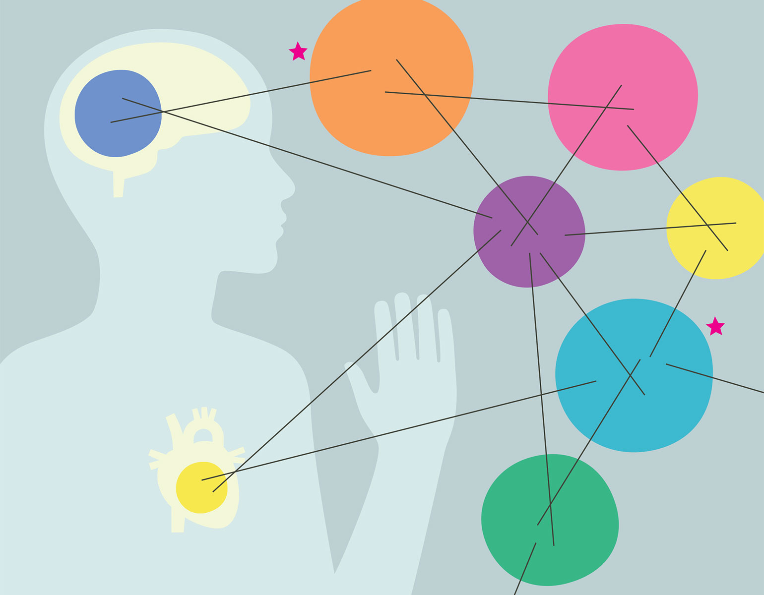
Were modern humans the first hominids capable of complex spoken language? If so, how did this unique capability evolve? New research by Robert B. Darnell and Erich D. Jarvis helps answer both of those questions, and could further our understanding of language and developmental disorders.

Darnell, who specializes in studying how RNA-binding proteins regulate gene expression, has spent more than three decades investigating a particular RNA-binding protein called NOVA1. NOVA1 is vital to brain development and neuromuscular control—Darnell has identified cases in which variations in NOVA1 are associated with developmental language and motor difficulties—and while it is found in animals ranging from mammals to birds, a particular variant of the protein, known as I197V, appears only in humans.

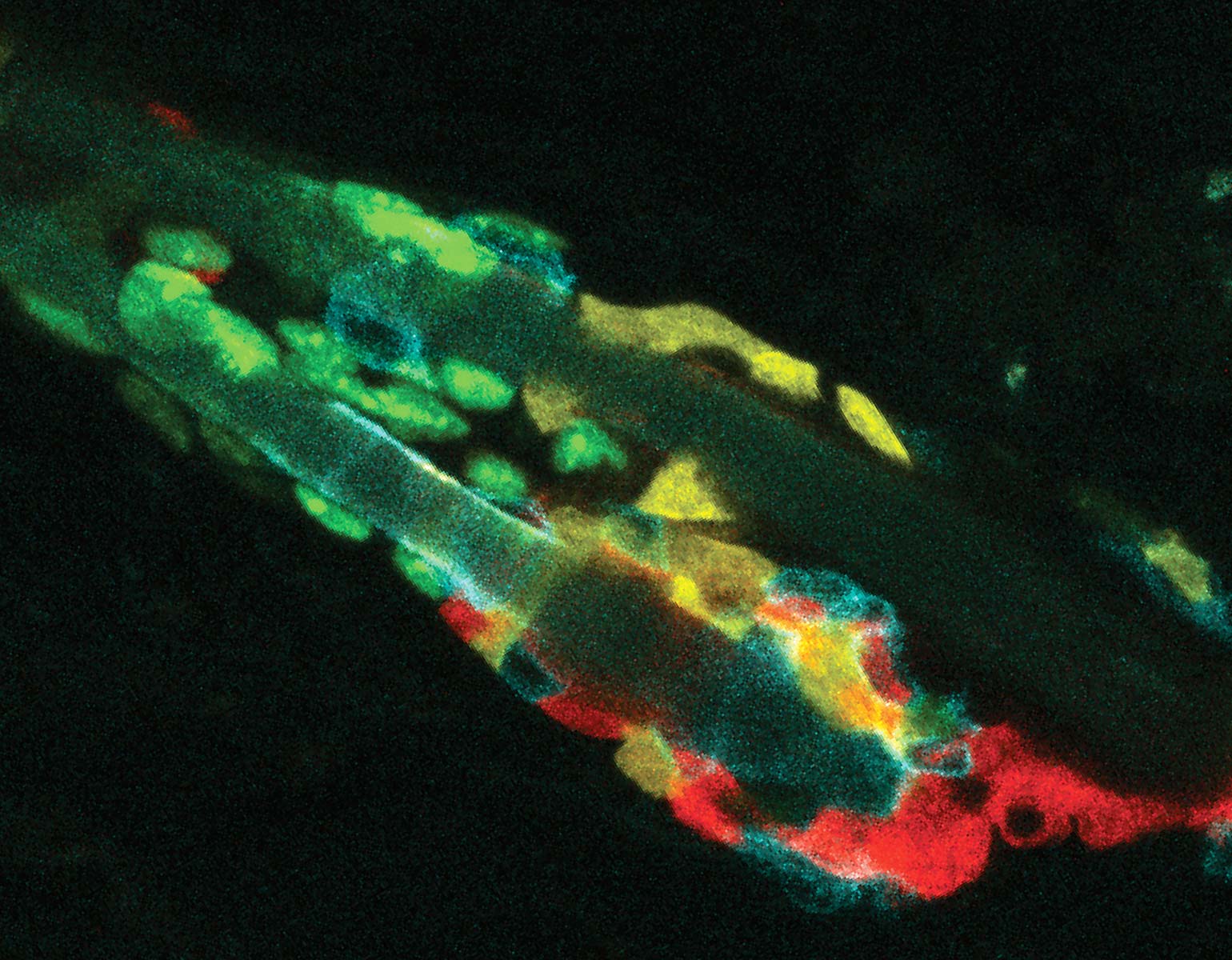
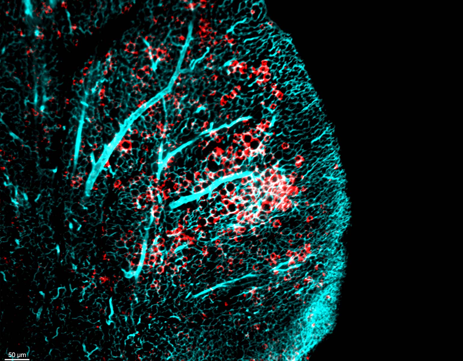
Yoko Tajima, a postdoc in Darnell’s lab, used CRISPR gene editing to replace the common NOVA1 protein found in mice with I197V. Intriguingly, the human-specific variant specifically affected RNA binding at sites related to vocalization.

Probing deeper, Darnell joined forces with Jarvis, who studies the molecular and genetic mechanisms underlying vocal learning. Over the next few years, the researchers documented altered vocal patterns among adult male mice and mouse pups of both sexes that carried the human variant.

“The single amino acid change in NOVA1 may make it a bona fide human ‘language gene,’” Darnell says. “Though certainly it’s only one of many human-specific genetic changes.”

To understand the potential influence of I197V on human evolution, the team compared the genomes of modern humans with those of our nearest relatives, the hominids known as Neanderthals and Denisovans. While these archaic relatives had the same version of NOVA1 found in nonhuman animals, the human-specific I197V variant was found in 650,052 of 650,058 modern human genomes analyzed, underscoring how it has become nearly ubiquitous, and suggesting it arose early in Homo sapiens’ evolution.

“Our data show that an ancestral population of modern humans in Africa evolved the human variant I197V, which then became dominant perhaps because it conferred advantages related to vocal communication,” Darnell says. “This population then left Africa and spread across the world.”

The team’s findings advance our understanding of when and how humans acquired their unique linguistic abilities. And by clarifying the role that NOVA1 plays in regulating language along with neural development and motor control, Darnell and Jarvis could also help scientists better understand a wide array of illnesses and impairments.

“Our discovery could have clinical relevance, ranging from children with language and developmental disorders to neuro­degenerative disease,” Darnell says.

A polinator’s oasis
Photo by Lori Chertoff

On Campus

A polinator’s oasis

Little bluestem, sea oats, butterfly weed, coneflowers, asters—Rockefeller’s lush campus is dotted with a host of colorful perennials. But these plants aren’t just beautiful; each one was carefully chosen by our landscaping team because it’s hearty enough to thrive in the campus’s fluctu­ating East River microclimate, where saline air and brisk winds can be tough on non-native species. Cultivating a space for this greenery to thrive among Manhattan’s concrete canyons means that bees, birds, and other wildlife can find a hospitable environment as well.

There’s more to CRISPR  than we knew
Laboratory of Bacteriology

Forefront

There’s more to CRISPR than we knew

Laboratory of Bacteriology

Bacteria have evolved powerful defenses against the viruses that prey on them. The most famous such defense, CRISPR-Cas9—a kind of molecular scissor that can snip away at viral DNA—was adapted to create the first FDA-approved genetic editing tool. But Luciano Marraffini, who helped identify CRISPR’s potential for genetic engineering, keeps finding more.

Most recently, Marraffini and his colleagues in the Laboratory of Bacteriology partnered with Dinshaw Patel at Memorial Sloan Kettering Cancer Center’s Structural Biology Program to study a class of molecules called CARF effectors that leap into action when bacteria are infected.

In the past year, the researchers have identified three CARF effectors that take different approaches to achieving the same goal: stymying viral propagation by bringing cellular activity to a grinding halt. Cad1 triggers a sort of molecular fumigation, flooding infected cells with toxic molecules. Cam1 slows their growth by altering their cell membranes. And Cat1 depletes a metabolite essential for cellular function, which cuts off the viral invader’s fuel supply.

“The range of both their enzymatic activities and structures is quite amazing,” says Marraffini, who adds that much remains to be learned about how these molecules work their antiviral magic. “It will be fascinating to see where this work leads us next.”

This enzyme heads off transcriptional catastrophe
Photo by Lori Chertoff

Forefront

This enzyme heads off transcriptional catastrophe

Photo by Lori Chertoff

Cells express their genetic instructions by transcribing DNA into RNA. Sometimes, though, that process goes dangerously awry, destabilizing the genome and contributing to a whole host of diseases.

A recent study from Seth Darst’s lab reveals how a particular enzyme helps prevent this kind of transcriptional catastrophe from occurring in bacteria—a finding that could inspire new strategies for targeting illnesses linked to genome instability.

All living things rely on an enzyme called RNA polymerase (RNAP) for transcription. While RNAP normally releases DNA after transcribing it, the enzyme sometimes remains clamped in place, reinitiating the process and creating potentially dangerous molecular structures called R-loops unless another enzyme called RapA intervenes.

Darst and colleagues demonstrated that the RapA enzyme functions as a bacterial Jaws of Life, prying open RNAP to stop it from inadvertently producing R-loops. Using advanced imaging techniques and a realistic substitute for bacterial DNA, the researchers captured the moment when RapA forces RNAP to let go of DNA. They also showed that E. coli bacteria engineered to lack RapA experienced genetic instability when stressed.

The findings suggest that RapA is a key safeguard against transcription-induced genome instability, and Darst suspects a similar mechanism may exist in all bacteria—and possibly across species.

“This work not only clarifies RapA’s role,” he says, “but also opens up broader questions about how all cells prevent transcription from becoming a genomic liability.”

How cancer can use lipids to hide
Laboratory of Metabolic Regulation and Genetics

Forefront

How cancer can use lipids to hide

Laboratory of Metabolic Regulation and Genetics

Some cancer cells are loud and proud, announcing their presence with chemical markers that allow the body’s immune system to find and destroy them. But others learn to hide, and a recent study from Kivanç Birsoy’s lab reveals that certain tumors rely on the fatty molecules known as lipids to do it.

One class of lipids stood out in particular: sphingolipids, which are named after the enigmatic Sphinx of Greek lore due to their initially puzzling structure and function. Scientists eventually came to view sphingolipids as important components of cell membranes—and useful fuel for hungry cancer cells. But Birsoy’s study, which was carried out in collaboration with Gabriel D. Victora’s lab, suggests that sphingolipids also play an active role in shielding cancer from immune detection.

“We believe modulating dietary lipids may be an interesting avenue to target cancer cells’ ability to evade immune cells.”

Cancer cells seemed to manipulate these lipids to distort the “eat me” signals that normally flag them for destruction. To test whether glycosphingolipids were essential for this deception, the researchers used an FDA-approved drug for Gaucher disease, a disorder in which lipids accumulate in certain organs, to block their synthesis. Sure enough, the drug dramatically slowed tumor growth in pancreatic, lung, and colorectal cancer models.

While more research is needed, the treatment appears to have worked by leaving the cancer cells exposed, suggesting that targeting sphingolipid production—through drugs or even lipids acquired through diet—could make cancers more vulnerable to immunotherapy.

“We believe modulating dietary lipids may be an interesting avenue to target cancer cells’ ability to evade immune cells,” Birsoy says.

Is metastasis a hereditary disorder?
Elizabeth And Vincent Meyer Laboratory Of Systems Cancer Biology

Forefront

Is metastasis a hereditary disorder?

Elizabeth And Vincent Meyer Laboratory Of Systems Cancer Biology

The vast majority of cancer deaths are caused by metastasis, or the spread of cancer cells from tumors to other parts of the body. Scientists long thought this was caused by mutations in the tumors themselves. Recent research by Sohail Tavazoie, however, indicates that metastasis is in part a hereditary disorder driven by our own DNA.

“We always thought that metastasis happens because people get a mutation in the tumor itself. But after searching tumors for decades, looking for a mutation that can explain metastasis, cancer biologists came up empty-handed. No one has ever found a real causal human mutation that promotes metastasis in the tumor,” Tavazoie says. “We’ve been so focused on the cancer cells, the ‘seeds,’ that we’ve ignored inherited genetic variations in otherwise healthy tissue—the ‘soil.’ It’s now clear that focusing on the soil is critical.”

Tavazoie’s team focused on PCSK9, a gene variant carried by roughly 70 percent of white women. Analyzing patient data from international cohorts and experimental data from mice, the team found that PCSK9 significantly increased the risk of metastasis within 15 years, raising it from 2 to 22 percent.

The work built on the lab’s previous research into skin cancer, which showed that variants of a gene called APOE caused metastasis by acting on a particular receptor. Interestingly, the PCSK9 variant appears to degrade that same receptor in breast cancer, triggering molecular changes that favor metastasis.

“What we’re seeing is that inherited genetics contribute to cancer metastasis by these two different cancer types,” Tavazoie says. “This makes me wonder whether the same pathway is related to the spread of other cancers as well.”

The results also point toward potential therapies. The team found preliminary evidence that an FDA-approved antibody that blocks PCSK9 and is currently prescribed for high cholesterol can suppress metastasis in lab models. In addition to seeking to test therapeutic targeting of this pathway with collaborators in the clinic, Tavazoie and his colleagues hope that future work will provide a path toward identifying those at highest risk of metastasis.

Mutations in 60 genes implicated in congenital heart disease
Illustration by Kari Modén

Forefront

Mutations in 60 genes implicated in congenital heart disease

Illustration by Kari Modén

Screening kids for the genes that cause congenital heart disease (CHD)—one of the most common birth defects and a leading cause of infant mortality—would be a game changer. But what if those same genes could tell you something about a child’s risk of neurodevelopmental problems as well?

That’s precisely what a sweeping study from the laboratory of Richard P. Lifton, who is Rockefeller’s president, promises to make possible. The study examined the genes of more than 11,000 children and identified mutations in 60 genes that are implicated in CHD, many of which are also linked to neurodevelopmental conditions such as autism. The results lend new insight into the biology of heart development and offer guidance for screening and early intervention across a wide array of disorders.

Although many of the mutations were spontaneous, the researchers were surprised to find that nearly half were inherited from parents who often showed no symptoms themselves. And while mutations in 33 genes were strongly associated with specific forms of CHD, others spanned a wide spectrum, producing narrow or broad cardiac outcomes depending on their exact nature. More than half of the implicated genes were also associated with neurodevelopmental disorders.

The findings have immediate clinical implications. Genetic screening may catch syndromes that would otherwise go undiagnosed, and early detection of neurodevelopmental risk could improve outcomes. Although every child in the study had already been diagnosed with congenital heart disease, genetic analysis revealed that nearly one third carried mutations linked to broader syndromes, many of which had gone unrecognized. In the absence of telltale symptoms, these additional conditions often escaped clinical detection, leaving associated cardiac or neurodevelopmental risks hidden in plain sight.

“This study sheds light on the complex architecture of CHD,” Lifton says. “With this information, physicians can better clarify diagnoses, anticipate outcomes, and assess the risk of CHD in future children.”

Balancing cooperation and competition
Illustration by Federica Bordoni

Forefront

Balancing cooperation and competition

Illustration by Federica Bordoni

If you thought courtship was tricky for humans, consider the games that fruit flies play.

Male fruit flies woo prospective mates by vibrating their tiny wings to produce high-frequency mating songs. But recent research from Vanessa Ruta’s lab reveals that competing males can “borrow” their rivals’ songs to win a female’s affections—or jam them with noise to spoil their chances.

Ruta’s team looked at what happened when two males competed for the attention of a single female. When one of the males was wingless and therefore incapable of singing—usually a recipe for courtship disaster—they found that it could sometimes sneak in a mating while its winged rival was singing, stealing his thunder and his mate.

“It turns out that mating success is not just about whether a male fly is the most vigorous in his courtship.”

Meanwhile, when both suitors had wings, they often tried to drown out one another’s songs with buzzy, high-pitched wing flicks. Further experiments confirmed that these flicks were indeed acts of sabotage that interfered with the female’s perception of courtship songs, activating brain pathways that blocked mating behavior.

The team also made neural recordings that revealed how male flies manage to balance this aggressive behavior with courtship.

Visual cues activate neurons associated with courtship, while the sound of a rival’s song switches on aggression-promoting neurons. Both neural systems can be co-activated, allowing rapid shifts between mating and aggressive interference. Genetically silencing aggression-related neurons eliminated the interfering wing flicks while leaving courtship behaviors intact, providing evidence of separate, interacting brain circuits.

The team’s findings highlight just how fluid and context-dependent fly behavior can be, while also underscoring the growing recognition in neuroscience that complex social behaviors, such as balancing competition and cooperation, don’t require complex brains—just precise, well-tuned neural circuits evolved for social survival.

“It turns out that mating success is not just about whether a male fly is the most vigorous in his courtship,” Ruta says. “It is also about whether he can successfully interweave courtship and aggression from moment to moment.”

New hope for reversing leptin resistance
Illustration by Sai Laung Linn

Forefront

New hope for reversing leptin resistance

Illustration by Sai Laung Linn

The modern weight-loss drugs known as GLP-1 agonists (think: Ozempic) have dramatically improved the health of millions. Yet we still haven’t solved the obesity crisis. Nor do we fully understand the one characteristic that 90 percent of obesity cases share: resistance to the hormone leptin. Recently, however, the lab of Jeffrey M. Friedman, who discovered leptin in the 1990s, revealed some of its molecular underpinnings—and a deeper understanding of this hormone which regulates eating.

Leptin is produced by fat cells and suppresses appetite in lean individuals. But in most obese individuals, this appetite-suppressing signal fails to register in the brain.

Earlier this year, Bowen Tan, Kristina Hedbacker, and other researchers in Friedman’s lab discovered a neural mechanism underlying leptin resistance: increased activity by a signaling molecule called mTOR in a particular population of neurons in the brain.

Intrigued, the researchers tested the effects of rapamycin, a drug that inhibits mTOR, on mice with diet-induced leptin resistance. The results were striking: “Obese mice fed a high-fat diet and treated with rapamycin lost significant amounts of weight,” says Tan.

“It essentially resensitized the animals to leptin,” Hedbacker adds. “Moreover, it was mostly fat that disappeared. That’s a significant difference from the effect of GLP-1 agonists, which cause the loss of both fat and muscle.”

In another study, Friedman’s lab identified a neural circuit that connects leptin to the jaw to stimulate chewing movements, suggesting that the impulse to eat may be more reflexive than previously thought. Inhibiting a specific group of neurons in the circuit led mice to consume more food and to make chewing motions even when food wasn’t nearby. Stimulating the same neurons, meanwhile, reduced both chewing motions and food intake, demonstrating an effective curb against hunger.

Together, these findings also bolster the idea that obesity is a far more complex condition than the old saying “calories in, calories out” might suggest.

“The available evidence tells us that obesity is an endocrine disorder, not a personal failing,” Friedman says. “It’s time for the stigma associated with obesity to end.”

What the gut tolerates
Photo by Lori Chertoff

Forefront

What the gut tolerates

Photo by Lori Chertoff

The gut is a gatekeeper, trained to recognize what belongs inside of us—and what doesn’t. But how does the intestinal immune system learn to distinguish friend from foe? And why does it sometimes make the wrong call, triggering a potentially dangerous allergic response to something as innocuous as a peanut or an egg?

“The big question is how we survive eating,” says Maria C.C. Canesso, a postdoc in the laboratory of Daniel Mucida. “Why do our bodies normally tolerate food, and what goes awry when we develop food allergies?”

A recent study led by Canesso and carried out in collaboration with the laboratory of Gabriel D. Victora offers clues. The researchers used new technology known as LIPSTIC, which catalogues cell-to-cell interactions, to identify how the intestinal lining teaches the immune system to tolerate dietary antigens, or the components of food molecules that immune cells recognize. Their findings reveal that two different intestinal immune cells capture food antigens and signal the immune system to stand down, preventing allergic reactions.

“Why do our bodies normally tolerate food, and what goes awry when we develop food allergies?”

The findings illuminate how the immune system maintains food tolerance. And while the scientists have not quite drawn a straight line from molecular mechanisms to food allergies, their work throws light on an intriguing path forward. If food allergies arise when intestinal cells lose their grip on immune balance, the authors suspect that we could one day fine-tune those cells to orchestrate tolerance rather than cause chaos.

Solving a  40-year-old  puzzle
Laboratory of Structural Biophysics and Mechanobiology

Forefront

Solving a 40-year-old puzzle

Laboratory of Structural Biophysics and Mechanobiology

A cell’s ability to move from place to place can be crucial. But that mobility can be a double-edged sword: The same mechanisms that allow an immune cell to rush to the site of an infection can also help metastatic cancer cells spread throughout the body. As such, new research from the lab of Gregory M. Alushin that reveals how cells get around could help improve cancer treatments—and even inspire new ones.

Many cell types have sensitive, finger-like protrusions called filopodia that help them move. But while filopodia require a certain amount of structural strength to enable locomotion, they are composed of highly floppy strands of protein known as actin filaments. These, in turn, must be bundled together by a protein called fascin in order to do anything useful.

“We’ve been able to detail essential design principles for the bundles.”

How fascin puts these bundles together has been a puzzle for more than 40 years—one Alushin’s team solved by developing advanced imaging technology that revealed the first clear three-dimensional images of fascin proteins binding actin filaments to form structures that “hit a sweet spot between strength and flexibility,” Alushin says.

The team’s insights could help improve drugs currently in development that stop cancer cells in their tracks by preventing fascin from bundling actin filaments into filopodia. Its findings could also lead to new therapies that work in a similar fashion.

“We’ve been able to detail essential design principles for the bundles, which could be really helpful information for finding new ways to interfere with their construction,” Alushin says.

Why cells sometimes eat their neighbors
Robin Chemers Neustein Laboratory of Mammalian Cell Biology and Development

Forefront

Why cells sometimes eat their neighbors

Robin Chemers Neustein Laboratory of Mammalian Cell Biology and Development

Every day, billions of our cells die and are swept away to make room for new ones. Some of these expired cells are literally devoured by specialized immune cells called phagocytes, which take their name from the Greek for “cell eater.” But others are consumed by their neighbors: non-phagocytes that normally do other jobs. So, how do these ordinary cells know when it’s time to suit up as temporary sanitation workers?

A recent study by Elaine Fuchs and her team provides answers. The researchers examined mouse hair follicles, which have relatively few phagocytes but still need to clear away dead cells to prevent unwanted inflammatory responses from occurring. They found that a pair of molecular sensors within follicle stem cells fuel the cyclical bouts of follicle and hair regeneration that (if you’re lucky) naturally occur throughout life. The sensors also pick up signals from their dying neighbors. In turn, this triggers living cells to eat the decaying ones, in turn extinguishing the signals and terminating the disposal operation before healthy cells get gobbled up too.

One of these sensors, called RXRα, detects the lipids secreted by dying cells, while the other, RARΥ, picks up the retinoic acid secreted by healthy cells. Dying cells trigger the cleanup process by releasing lipids, and once all the dead cells have been eliminated, only the retinoic acid signal from the healthy cells remains, shutting the program down because the two work together to unleash the phagocytic process. “It’s a really beautiful way to keep the area clean,” says Katherine Stewart, a former research associate in Fuchs’s lab.

The team’s findings have implications that go far beyond hair follicles, however. For example, stem cells in parts of the brain, breasts, and lungs also moonlight as ersatz phagocytes, keeping their own neighborhoods free from unwanted debris.

“For our body’s stem cells, this may be their way of keeping tissues fit by clearing out naturally dying cells and guarding against inflammation,” Fuchs says.

Metabolic havoc
Weslie R. and William H. Janeway Laboratory of Molecular Metabolism

Snapshot

Metabolic havoc

Weslie R. and William H. Janeway Laboratory of Molecular Metabolism

As body weight fluctuates, fat cells—collectively known as adipose tissue—wax and wane. This oscillation can wreak havoc on the body’s metabolism, especially in the case of yo-yo dieting, a phenomenon that’s been tied to a higher risk of developing diabetes, fatty liver disease, and
hypertension.

Mascha Koenen, a postdoc in Paul Cohen’s lab, suspects that’s largely a consequence of how yo-yo dieting feeds chronic inflammation simmering in visceral adipose tissue, a type of fat stored deep in our abdomen that contains a complex web of blood vessels and immune cells.

To probe deeper, Koenen took tissue samples from obese mice that went through a round of extreme weight loss, and stained their white blood cells—known as macrophages—red. The tiny crimson, crown-like structures that emerged revealed an unexpected pattern of sustained inflammation after weight loss. Koenen is now tracking how these immune cells interact with other cell types—whether they are essential to resetting the fat tissue to a normal state, or, alternatively, if they play a role in the detrimental outcomes associated with weight cycling. She hopes her research will inspire new efforts to treat the comorbidities that can linger long after the dieting is over.

Listening in
Photo by Chris Taggart

Gadget

Listening in

Photo by Chris Taggart

Say you’re standing in a loud, crowded cocktail party: Somehow, you find yourself effortlessly concentrating on the one conversation you’re participating in. For this deceptively mundane feat, you can thank your cochlear amplifier—rows of sensory hair cells deep within the inner ear that create tiny bursts of mechanical energy to strengthen faint sounds and sharpen tones before they reach the brain.

When this amplifier fails—whether through aging or noise exposure, for example—clarity fades and hearing loss begins. This tiny piece of biology has been maddeningly hard to study: The cochlea sits behind one of the densest bones in the body and is far too delicate to withstand probing in vitro. And until scientists can see the cochlear amplifier’s cellular gears in action, they can’t fully grasp how we process sound, why the system breaks down, or how to build hearing aids that might rival the ear’s natural finesse.

That’s why Rodrigo Alonso and Francesco Gianoli from the Hudspeth lab, working with instrumentation engineer Nicholas Belenko from Rockefeller’s Gruss Lipper Precision Instrumentation Technologies Center, created a way to keep cochlear amplifiers alive and functioning outside the body. By meticulously controlling ion balance, oxygen, temperature, and pressure, they can preserve the amplifier’s native environment, giving scientists unprecendented experimental access to its cellular components.

Researchers are now using this new system to study how mammalian inner ear cells behave across the full range of audible frequencies, measuring motions smaller than a billionth of a meter.

“For the first time, we can keep a tiny slice of the inner ear alive in the lab and watch the ear’s built-in amplifier at work under controlled conditions,” says Gianoli. “By pinpointing the key elements that give this system its sensitivity, we can see exactly where mutations, diseases, noise, and aging start to erode it.”