The brain cells most vulnerable to Huntington’s
Laboratory of Molecular Biology

Forefront

The brain cells most vulnerable to Huntington’s

Laboratory of Molecular Biology

Scientists know a good deal about Huntington’s disease, an inherited neurodegenerative disorder that slowly robs patients of their physical and mental health.

They know, for example, that it is caused by mutations to a particular gene; that these mutations involve the excessive repetition of tiny stretches of DNA bases known as CAG repeats; and that these repeats corrupt the proteins that the gene produces, ultimately causing neurons in certain parts of the brain to die.

But they have not yet learned enough about the molecular mechanisms underlying this process to have developed drugs that can stop or reverse the disease.

Hence the excitement when researchers in Nathaniel Heintz’s Laboratory of Molecular Biology recently teased out some hitherto unknown nuances of those mechanisms, providing potential targets for therapeutic interventions.

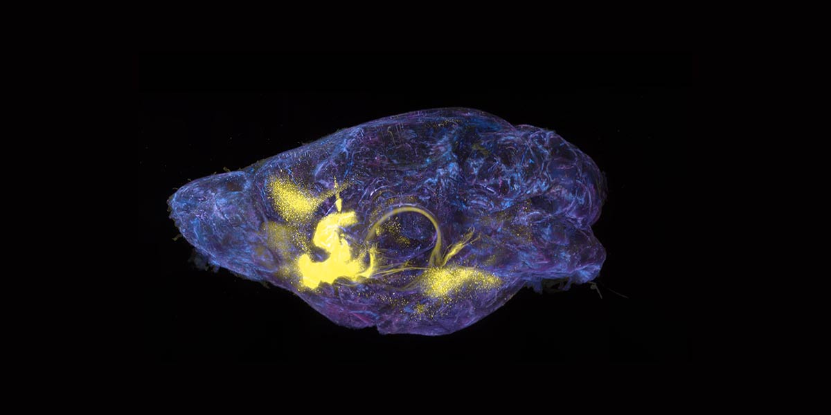
Using cutting-edge molecular profiling techniques, the scientists discovered that CAG repeats are unstable—and therefore likely to produce more toxic proteins—in only certain types of brain cells. They also found that other cell types proved surprisingly resilient to the repeats.

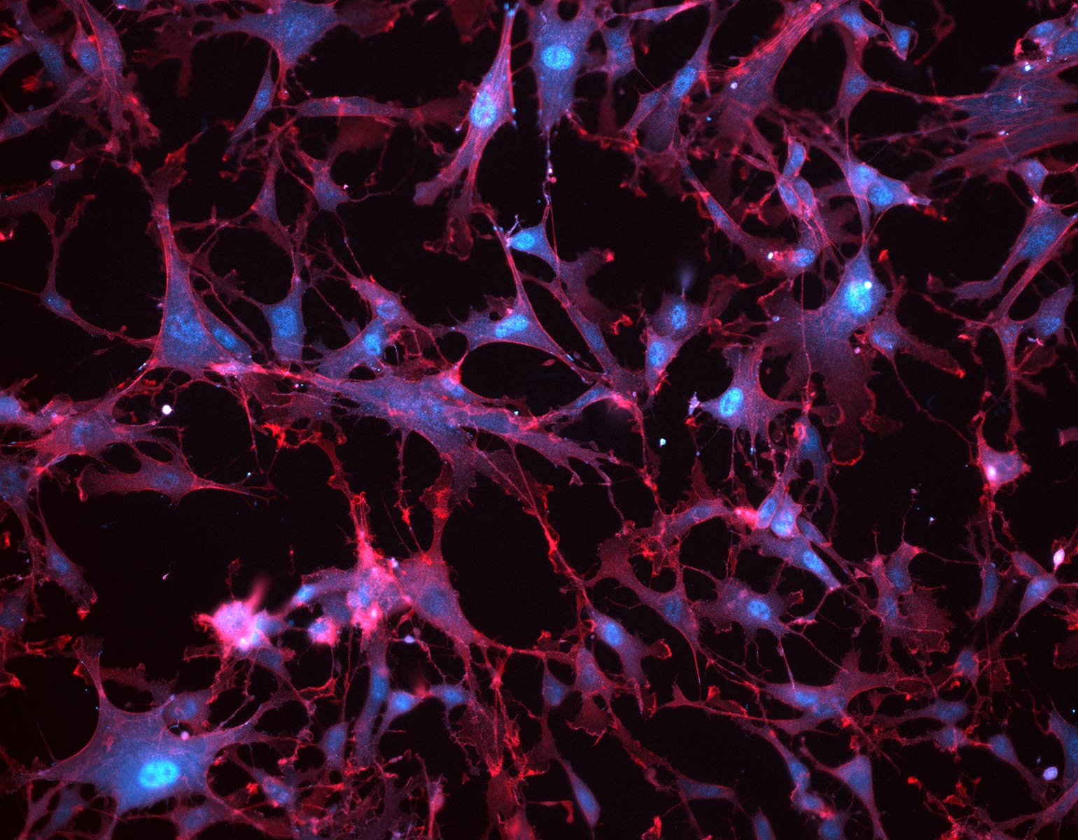
In separate but complementary studies from Heintz’s lab, Kert Mätlik, a research associate, and Christina Pressl, an instructor in clinical investigation, examined the brain cells of people who had died from Huntington’s. The scientists focused on two brain regions that are profoundly affected by the disease: the striatum and the cortex.

In the striatum, Mätlik found that CAG repeats were particularly unstable in medium spiny neurons, the cells most likely to be lost during the progression of Huntington’s. Moreover, in their nuclei these neurons contained high levels of a protein complex called MutSβ that is known to promote expansion of CAG repeats in experimental models.

Meanwhile, Pressl discovered that although different types of pyramidal neurons found in the deep layers of the cortex had very long CAG repeats, only one population was more likely to die. The discovery “puts another cell type on the map for increased vulnerability to the disease,” she says.

Her study also provided evidence that the susceptible neurons in the cortex project slender fibers called axons into the striatum, where Mätlik’s medium spiny neurons reside; communication between the two brain regions is already known to falter in Huntington’s. As such, the team’s combined findings suggest that the vulnerable cells in both areas may be connected. “When it comes to Huntington’s, the entire neural network breaks down at some point,” Pressl says.

Heintz, the James and Marilyn Simons Professor, hopes to build on these studies to answer more questions about the disease.

“Is there a specific length of repeats at which the cells become dysfunctional? At what CAG repeat length do the cells die, and does it differ depending on the cell type?” he asks.

“We need to understand these things in order to develop new treatments for this devastating disease.”
History books
Photo by Lori Chertoff

On Campus

History books

Tucked behind the circulation desk inside Rockefeller’s Rita and Frits Markus Library, readers can find Rockefeller student dissertations dating back to 1959, the year of the university’s first convocation. On this storied bookcase sits early work that developed into biomedical breakthroughs, from revealing the chemical structure of antibodies to demystifying the circadian clock to explicating how humans regulate body weight. Among the authors are now two Nobel laureates, 27 members of the National Academy of Medicine, and 36 members of the National Academy of Sciences, along with researchers who have achieved countless other noteworthy distinctions.

First glimpses of the human form
Laboratory of Synthetic Embryology

Forefront

First glimpses of the human form

Laboratory of Synthetic Embryology

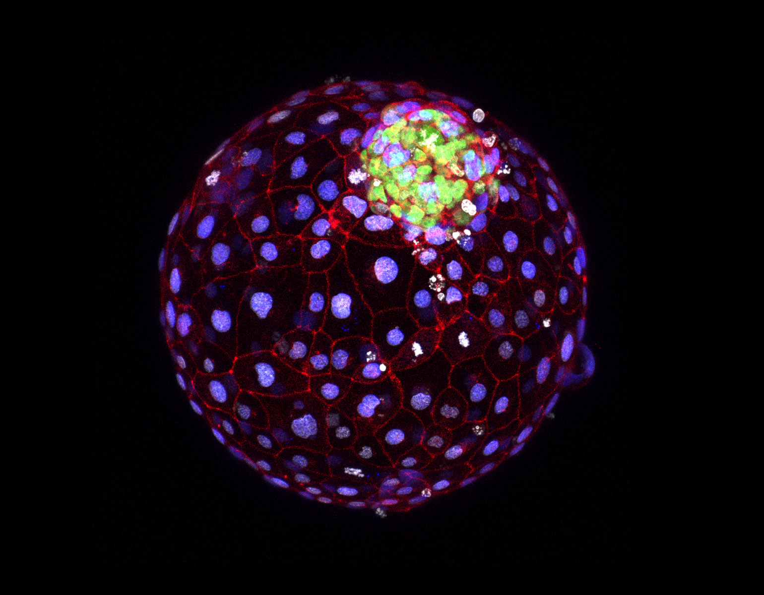
Mere weeks into human embryonic development, an indistinct ball of cells called a blastocyst rearranges itself into an orderly three-layered structure—a process called gastrulation that sets up the eventual emergence of the human form.

Understanding the molecular underpinnings of this pivotal event could help scientists prevent miscarriages and head off a host of serious disorders. But gastrulation has long been a black box. “It is the first moment that separates our heads from our behinds, and yet we had never seen ourselves at that stage,” says Ali H. Brivanlou, the Robert and Harriet Heilbrunn Professor.

That changed this year when Brivanlou and his Laboratory of Synthetic Embryology created a new platform for studying human gastrulation through the use of blastoids—models of blastocysts developed from stem cells that can be cloned, manipulated, and programmed in the lab. Crucially, the platform allows blastoids to attach to plastic surfaces, much as real blastocysts attach to the uterus.

1998

Data

Year the first human stem cell line was created.

Thanks to this innovation, the team was able to directly observe key moments in process, including the emergence of molecular markers for two crucial structures in embryonic development: the so-called primitive streak, which marks the beginning of gastrulation; and the mesoderm, which gives rise to muscles, bones, and the circulatory system.

The future applications are many, says first author Riccardo De Santis, a research associate in Brivanlou’s lab: “A better understanding of gastrulation impacts everything from survival of the fetus to autism to neurodegeneration.”

Taking a strategic pause
Illustration by Sebastian Thibault

Forefront

Taking a strategic pause

Illustration by Sebastian Thibault

Mycobacterium tuberculosis (Mtb) is a wily foe, adept at bobbing and weaving around the immune system and antibiotics alike, and sometimes lying dormant for years.

“It’s a very smart bacterium, with a lot of tricks,” says Shixin Liu, head of the Laboratory of Nanoscale Biophysics and Biochemistry.

Liu has pulled the curtain back on one such trick, working with Jeremy M. Rock and Corinne P. Greenberg Women & Science Professor Elizabeth Campbell, who heads the Laboratory of Molecular Pathogenesis. Together, they found that Mtb can control its gene expression by pausing transcription, or the process by which DNA is copied to RNA. Taking transcriptional “breathers” may give the bacterium a chance to adapt to changing conditions, such as a different environment or the presence of antibiotics.

13 million

Data

Number of people living with inactive TB in the U.S., according to CDC estimates.

Previously, bacteria weren’t thought to prominently use such strategic pausing to regulate their gene expression, which is common in more complex organisms. “The findings were so unexpected that I initially didn’t believe them,” says Rock, the Penrhyn E. Cook Associate Professor and head of the Laboratory of Host-Pathogen Biology.

Their startling discovery came via new technology: SEnd-seq, a high-resolution RNA sequencing tool developed by Liu and Xiangwu Ju, a senior research associate in the Liu lab, which the team used to analyze Mtb’s RNA transcripts at an unprecedented level of detail.

The researchers suspect that homing in on the pausing mechanism could provide a particularly fruitful avenue for identifying new drug targets against their cunning bacterial adversary.

“We can use this information to think about how we might inhibit its life cycle,” Liu says.

Sensory nerves help cancer spread
The tumor on the right with few nerves remains localized; the one on the left with many more has spread. Laboratory of Systems Cancer Biology

Forefront

Sensory nerves help cancer spread

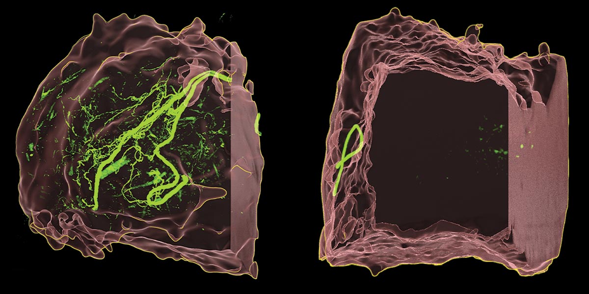
The tumor on the right with few nerves remains localized; the one on the left with many more has spread. Laboratory of Systems Cancer Biology

cancer has an unsettling ability to circumvent our natural defenses, growing and metastasizing from one place to another despite the body’s best efforts to contain it. Now, Rockefeller researchers have shown how the disease appears to be co-opting the nervous system to extend its reach.

Previous studies had shown that cancer and the nervous system often pair up in a malignant pas de deux known as the “neuro-cancer axis,” with tumor cells recruiting nerves to their primary site and the nervous system kickstarting tumor growth in turn.

But researchers in Sohail Tavazoie’s Elizabeth and Vincent Meyer Laboratory of Systems Cancer Biology have learned that nerves don’t just foster tumor growth, they also drive metastasis, which is the main cause of death in most cancers. “This is an exciting discovery—no one has seen peripheral nerves release a signal to enhance metastasis before,” explains Veena Padmanaban, a postdoctoral fellow in the Tavazoie lab and lead author of the study exploring the relationship between sensory nerves and breast cancer.

Tavazoie’s team used mouse models to compare innervation between highly metastatic and less metastatic tumors and found that sensory nerves were spurring breast cancer metastasis. When they analyzed publicly available data, they discovered a similar trend among human breast cancer patients. But the implications of their work could be even broader: The group found that nerve-tumor interactions also activated genes in nearby cells through a process known as RNA signaling.

While the ramifications of that aren’t yet clear, “it was unexpected and may have relevance that extends beyond cancer,” Tavazoie, Leon Hess Professor, explains.

In the meantime, the team managed to impede the growth and metastasis of multiple models of breast cancer when they treated mice with aprepitant, an FDA-approved anti-nausea medication already commonly prescribed for chemotherapy patients.

“Because aprepitant is already approved and safe, oncologists may consider clinical trials to test the impact of this medication on cancer progression in patients with breast cancer,” Tavazoie says.

Getting the message
Photo by Lori Chertoff

Forefront

Getting the message

Photo by Lori Chertoff

Despite recent efforts to catalog all the different cell types in our bodies, the great mystery of how those cells work together to form tissues and organs remains unsolved. A comprehensive map detailing how cells communicate and collaborate would revolutionize basic biology. But no such “interactome” is possible without a method of reliably tracking millions of intercellular interactions.

Gabriel D. Victora’s Laboratory of Lymphocyte Dynamics developed a limited version of such a method several years ago. The team named its approach LIPSTIC because it involves labeling cellular structures that touch when two cells make fleeting, “kiss-and-run” contact before parting ways. In essence, it allows researchers to track physical cell-to-cell interactions by causing any cell that “kisses” another to leave behind a biochemical mark like a lipstick trace, which can later be analyzed in the lab. The platform was initially designed only to record a specific type of interaction between immune cells, but, when other researchers got wind of LIPSTIC, they began clamoring for a universal version.

“With uLIPSTIC we can ask how cells work together, how they communicate, and what messages they transfer.”

Hence the new and improved tool, which allows scientists to theoretically smear LIPSTIC on any kind of cell and track its physical interactions with other cells. Laurie and Peter Grauer Professor Victora and his team hope to use uLIPSTIC to better understand how cells unite into tissue at the molecular level, and they envision it serving as a central tool for building a comprehensive cell-to-cell interactome.

“With uLIPSTIC we can ask how cells work together, how they communicate, and what messages they transfer,” Victora says. “That’s where biology resides.”

Rewiring the brain for addiction
Laboratory of Molecular Genetics

Forefront

Rewiring the brain for addiction

Laboratory of Molecular Genetics

Scientists have long understood that addictive drugs like cocaine and morphine hijack the brain’s internal reward system, causing us to crave them while simultaneously disrupting our natural urges to eat and drink. Yet exactly how these drugs rewire our brains hasn’t been understood. A collaboration between scientists at Rockefeller and Mount Sinai recently shed considerable light on the situation by identifying a common reward pathway in a brain region involved in pleasure, motivation, and decision-making. Known as the nucleus accumbens (NAc), this region works closely with dopamine and serotonin, which modulate mood and reinforce behaviors that feel good, such as eating a meal, socializing—or using drugs.

“What we hadn’t been able to understand is how repeated exposure to drugs alters the function of these neurons, resulting in escalated drug-seeking behaviors and a shift away from normal drives such as eating and drinking,” says Bowen Tan, lead author on the paper and a graduate student in the Laboratory of Molecular Genetics headed by Jeffrey M. Friedman.

“What we hadn’t been able to understand is how repeated exposure to drugs alters the function of these neurons.”

To capture what’s really happening inside the brain when it’s exposed to both drugs and natural rewards, Marilyn M. Simpson Professor Friedman and his team turned to advanced brain imaging techniques developed in the Laboratory of Neurotechnology and Biophysics, headed by Alipasha Vaziri, who worked with them to track the neural activity of mice in real time. Coupling those high-tech imaging methods with cutting-edge molecular and genomic techniques, the researchers identified for the first time how drug addiction warps natural urges by commandeering a molecular pathway that plays a crucial role in neural plasticity, the process that neurons use to reinforce learning and memory.

A gene called Rheb is at the center of this pathway. When drugs like cocaine activate brain cells expressing Rheb, this stimulates a pathway that appears to alter how neurons process stimuli from food and water. This may explain why mice and humans who are addicted to these substances escalate drug use and show decreased needs for natural rewards like food and water.

The team’s findings provide new insights about the different effects of natural rewards and addictive drugs, potentially paving the way for new approaches that more directly address how addiction subverts expected behavior.

How a key gene contributes to autism
Laboratory of Developmental Neurobiology

Forefront

How a key gene contributes to autism

Laboratory of Developmental Neurobiology

autism spectrum disorder (ASD) has a strong genetic component. But sorting out how the scores of genes that have been linked to the developmental disorder actually contribute to it has been difficult. Recent research by Mary E. Hatten is bringing some much-needed clarity to the situation.

In 2018, Hatten and her colleagues in the Laboratory of Developmental Neurobiology discovered how defects in the protein produced by a gene called ASTN2 disrupted circuitry in the cerebellum of children with ASD and other neurodevelopmental conditions. This year, Hatten’s lab found that knocking out the same gene in mice led to several hallmark ASD behaviors, as well as to physiological changes in the brain.

Mice that lacked ASTN2 vocalized and socialized less but were more hyperactive and repetitive in their behavior than their wild-type nestmates. When briefly isolated, for example, knockout pups were less likely to call out for their mothers and used simpler vocalizations.

Similar communication and behavior issues are common in people with ASD, says first author Michalina Hanzel: “Some autistic people don’t understand metaphor, while others echo language they’ve overheard, and still others do not speak at all.”

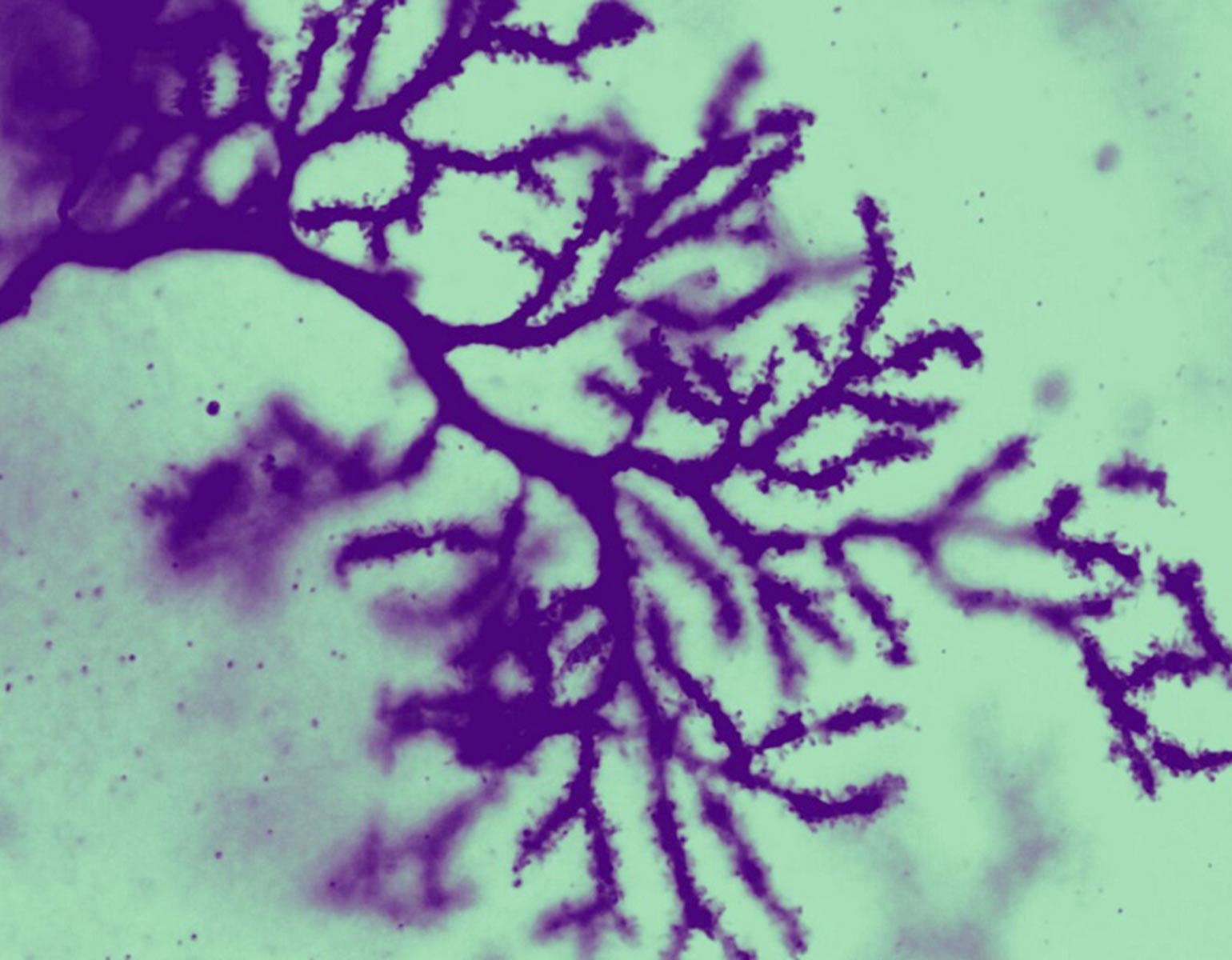
The physiological changes centered around abnormalities in brain cells that are located in specific parts of the cerebellum.

“The differences are quite subtle, but they are clearly affecting how the mice are behaving,” says Hatten, the Frederick P. Rose Professor. “The changes are probably altering the communication between the cerebellum and the rest of the brain.”

In addition to providing crucial insight into the genetic causes of ASD, the team’s findings add to the growing body of evidence that the cerebellum—the oldest cortical structure in the brain—is important not just for motor control but also for language, cognition, and social behavior. “It’s a big finding in the field of neuroscience,” Hatten says.

Tiny and mighty
Photo by Lori Chertoff

Forefront

Tiny and mighty

Photo by Lori Chertoff

It’s hard to study how brain activity correlates with behavior when the animals being studied aren’t behaving freely. This poses a unique challenge for neuroscientists, since tracking activity across the brain of a moving, active subject is no mean feat.

The solution is generally a head-mounted microscope that mammals wear like a hat. But mice, the go-to model organisms for understanding the brain at work, cannot carry anything heavier than a penny. And microscopes that small can only capture what’s going on in a tiny portion of the brain.

“Those microscopes typically support rather small imaging fields of view,” says Alipasha Vaziri, head of the Laboratory of Neurotechnology and Biophysics, who decided to tackle this issue from a unique angle.

Using conventional methods to increase the field of view had required a frustrating trade-off: Either reduce resolution to widen the field of view or maintain single-cell level resolution by adding more complex and multi-element lenses, which also adds an unwieldy amount of weight.

Vaziri circumvented the problem by going back to the drawing board and coming up with a radical solution: What if the lenses weren’t actually necessary? If an optical system can faithfully encode 3D points from a sample to 2D points on a camera, that’s sufficient, since that process can be computationally reversed. That is, the optical system does not necessarily need to be image-forming, per se. The result is an innovative, essentially lensless, microscope that’s light enough for a mouse to wear on the go, yet packs a field of view wide enough to get the job done (pictured here, not actual size).

“This tech could advance our understanding of how brain-wide distributed neuroactivity relates to naturalistic behavior,” Vaziri says.

Rewriting the textbooks on telomeres

Forefront

Rewriting the textbooks on telomeres

Like habitable planets orbiting distant stars, the chromosomes that contain our DNA are subject to a Goldilocks principle. Telomeres, the protective caps found at chromosome ends, must be kept at just the right length: too short, and dangerously accelerated aging ensues; too long, and they predispose their owners to cancer. Precise regulation is paramount, and Titia de Lange’s Laboratory of Cell Biology and Genetics is cracking the code on how three specific enzymes keep telomeres in check.

50-70

Data

Typical number of times a human cell can divide before its telomeres become too short to operate properly.

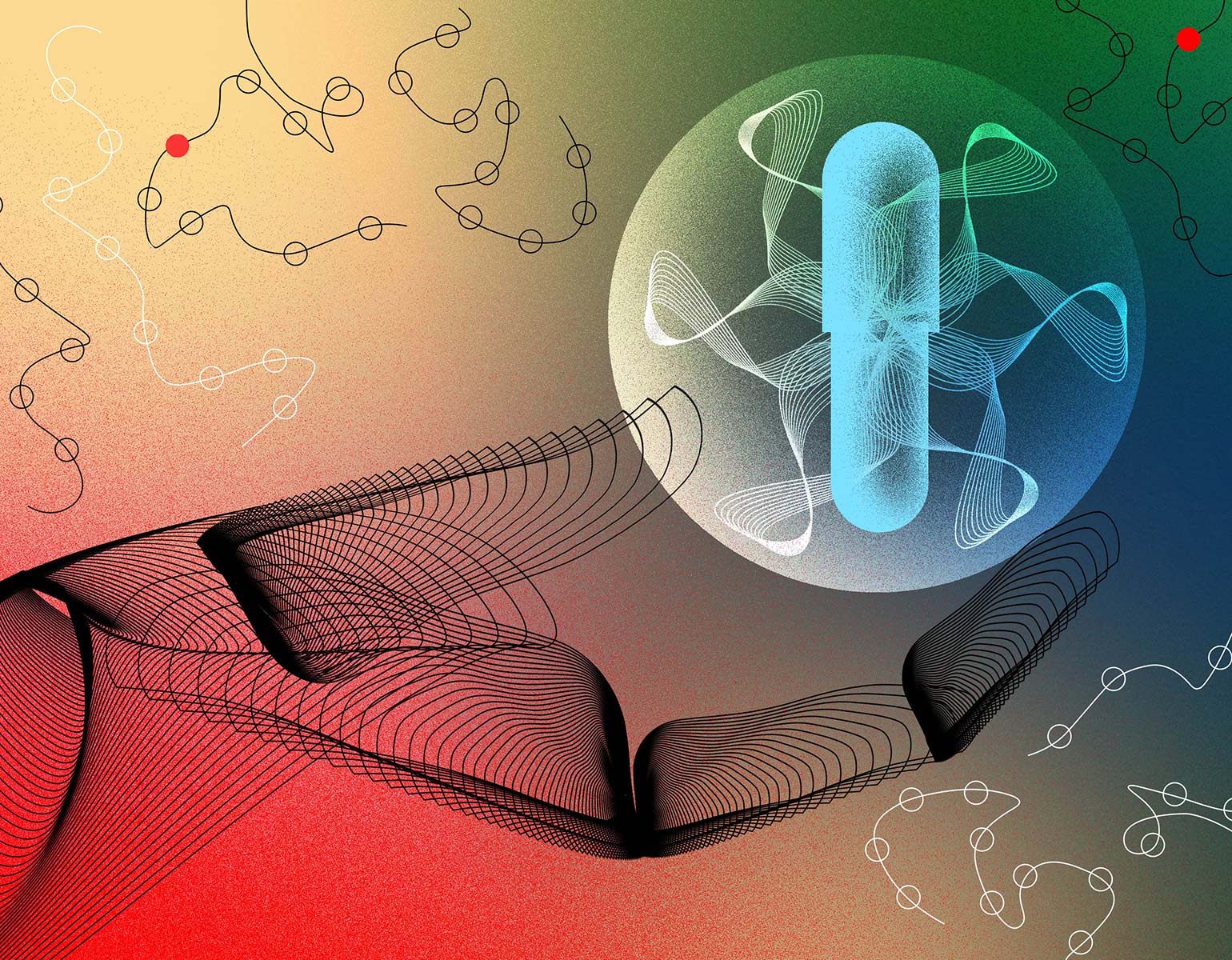
Among their many findings, de Lange’s team discovered that two of these enzymes work in concert to ensure proper telomere maintenance. The telomerase enzyme helps prevent telomeres from growing shorter with each round of DNA replication. But every DNA double helix has two strands that must be copied, and telomerase is capable of handling only one of them. In a recent study, Leon Hess Professor de Lange and her team demonstrated that a second enzyme—known as the CST–Polα-primase complex—is responsible for managing the other strand. They further determined how that enzyme complex is recruited and regulated.

In addition to fundamentally changing our understanding of telomere biology, these insights could lead to potential treatments for individuals who suffer from telomere disorders such as Coats plus syndrome, a devastating multi-organ disease characterized by abnormalities in the eyes, brain, bones, and GI tract.

“Telomerase should only be acting at the natural ends of healthy chromosomes. When it goes to work anywhere else, very bad things happen.”

Meanwhile, related work from de Lange’s lab elucidated a key mechanism for preventing telomerase from accidentally preserving damaged DNA. The main actor turned out to be the enzyme ATR kinase, which stops telomerase from adding telomeres to the ends of broken bits of DNA rather than to the ends of intact chromosomes, where they belong. In light of many studies suggesting that telomerase runs amok in tumors, de Lange is now exploring whether glitches in this self-protective process may help maintain the altered chromosomes that promote cancer progression.

“Telomerase should only be acting at the natural ends of healthy chromosomes,” de Lange says. “When it goes to work anywhere else, very bad things happen.”

Passing the test
Illustration by Brian Stauffer

Forefront

Passing the test

Illustration by Brian Stauffer

Deep-learning algorithms can unpack the unique 3D structures of a protein in less than an hour—a task that used to take scientists a year. But do these models have the same ability to guide drug discovery as experimental structures do?

If so, the implications for medicine would be immense: Once the molecular nuances of a protein’s structure have been identified, researchers could begin targeting it with drugs to correct dysfunctions, combat infections, and improve health.

Research by Jiankun Lyu indicates that at least one such algorithm, Google DeepMind’s AlphaFold2, is up to the job. Lyu’s lab used sophisticated molecular docking (or virtual screening) software to sift through billions of chemical compounds—searching for potential drugs by matching them against protein structures—and found that for the two drugs they tested, AF2’s predicted structures are just as capable of guiding virtual drug screening as are experimental structures. Based on those findings, Lyu says, the algorithm could potentially expedite some drug-discovery projects by as much as several years.

Future deep-learning algorithms will almost certainly be even more powerful. An exciting prospect, but Lyu advises an equal measure of caution: “A lot of AI is currently overpromised and under-delivered. If we don’t tread carefully, AI in biomedicine will end up being just another hype.”

Case in point: Google DeepMind recently released AlphaFold3, which Lyu describes as “a huge upgrade” over its predecessor. Unlike AlphaFold2, however, AlphaFold3 is a black box; the company has not released the underlying deep-learning model to researchers, which means that they can’t properly test it.

“We would not have been able to run the current study on AlphaFold3,” says Lyu, now head of the Evnin Family Laboratory of Computational Molecular Discovery. “And without that, we can’t know whether the new model is better for drug discovery.”

Cellular sensation
Laboratory of Structural Biophysics and Mechanobiology

Snapshot

Cellular sensation

Laboratory of Structural Biophysics and Mechanobiology

Any New Yorker jostling onto the subway during the morning rush doesn’t need eyes to know the train is crowded. Likewise, those riding home later at night can sense the relative quiet. So too does every cell in the body detect its environs and respond appropriately through a technique scientists call mechanosensation.

Whether a cell’s habitat is soft and spacious, or hard and crowded, has a powerful effect on gene expression, spurring different cellular actions. To get that information, eukaryotic cells (the type humans are made of) send out feelers connecting the nucleus to the cell’s microenvironment, via a network of specialized proteins.

Donovan Phua, a graduate student in Gregory M. Alushin’s Laboratory of Structural Biophysics and Mechanobiology, wants to know exactly what those sensors (like FLH2, seen here in cyan) are up to inside the nucleus, the headquarters for genes. Understanding that helps explain how mechanical forces act on normal cells—as well as the bad behavior of diseased ones, which create tumors by growing uncontrollably despite dense surroundings. Phua hopes that this work could inform efforts toward new cancer therapies that teach rogue cells their proper place.

1,000+ channels
Photo by Lori Chertoff

Gadget

1,000+ channels

Photo by Lori Chertoff

The mass spectrometer was invented in 1912 by English scientist J.J. Thomson (famed discoverer of the electron). Ever since, it’s been a lab standard, the go-to tool for deducing the chemical composition of scientific samples.

The problem is, as it became more sophisticated, it also became more cumbersome. “Instead of doing everything simultaneously, as early machines could when the whole process was simpler, each step must now be completed one after another,” says Brian T. Chait, Rockefeller’s Camille and Henry Dreyfus Professor and head of the Laboratory of Mass Spectrometry and Gaseous Ion Chemistry. Thus: First the mass spec ionizes a sample; then it determines that sample’s mass-to-charge ratio; finally it measures the outcome. This costs scientists precious time, because each run through the apparatus accommodates only a minuscule fraction of a given sample, which are often precious commodities, like donated cancer cells. “Mathematically, it’s the equivalent of trawling Niagara Falls, looking for rare species of a teeny fish with a tiny bucket.”

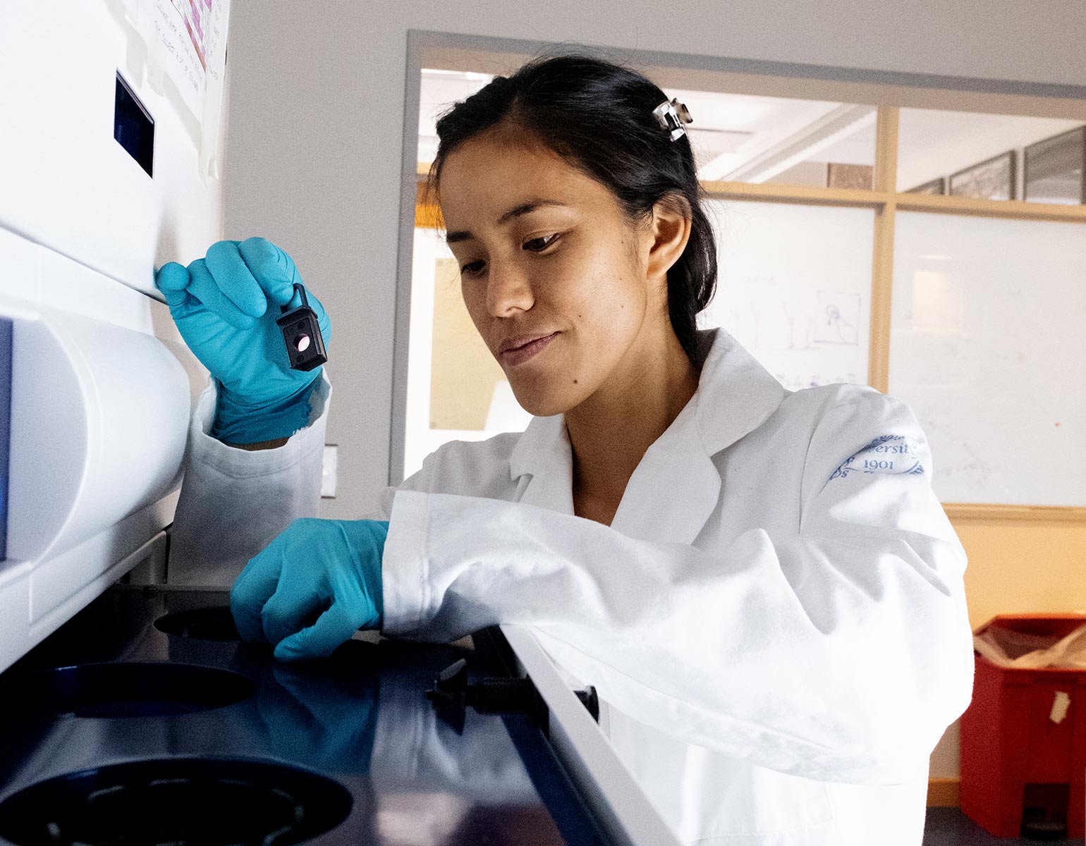
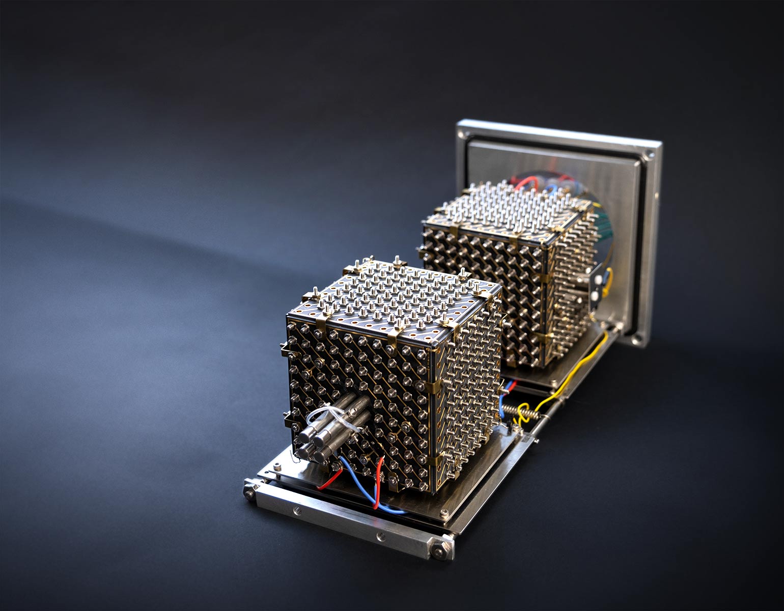
So, Chait and Andrew Krutchinsky, a senior research associate in Chait’s lab, developed a tool that reintroduces the concept of parallel analysis, potentially increasing the mass spec’s processing capacity 1,000-fold. Rather than blasting samples through one at a time, their prototype—a cubic device hand soldered, assembled, and retrofitted by the duo—adds 1,000 channels for weighing and sorting molecules in parallel.

Chait and Krutchinsky can’t wait to work faster. It took Chait and collaborator Michael P. Rout, Rockefeller’s George and Ruby DeStevens Professor, 30 years to piece together a comprehensive, 3D model of the nuclear pore complex—a cellular gatekeeper that’s implicated in diseases as diverse as cancer and Alzheimer’s. “We needed an expediting tool, because we want to see more of how this pore behaves in action,” says Chait. “Plus, there are thousands of other biomolecular machines we have yet to explore.”