Interview
A new way to look at neurons
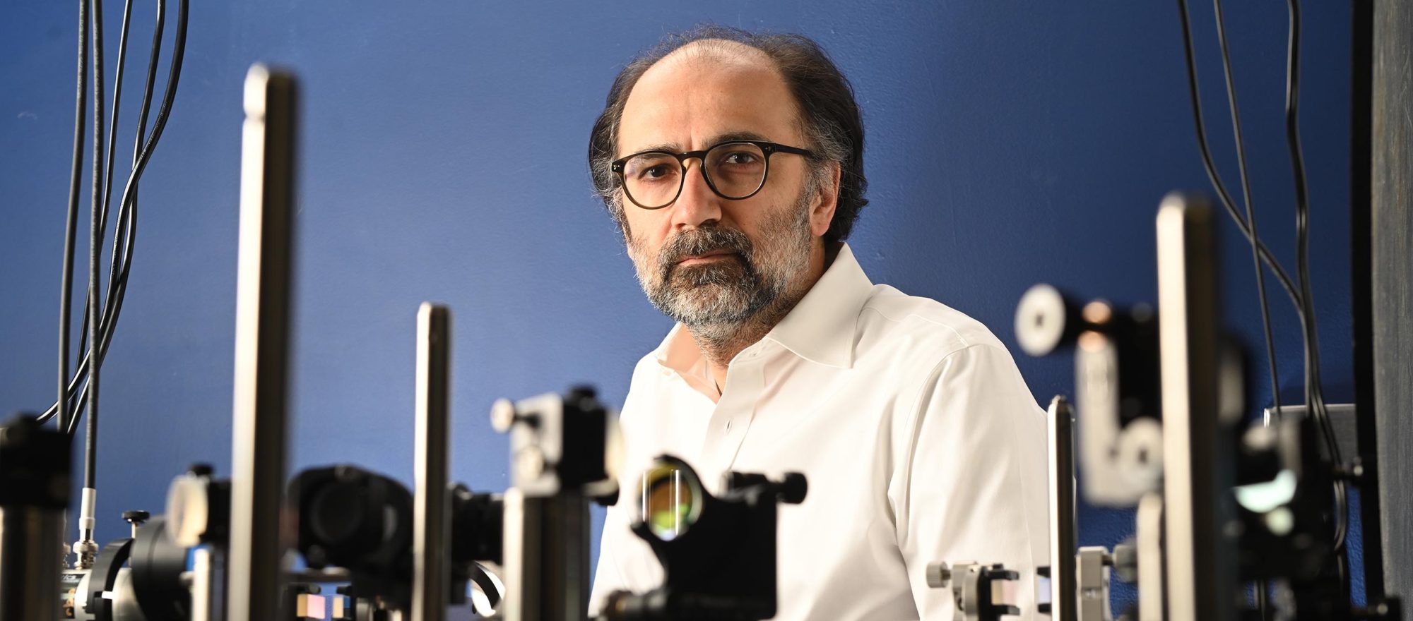
with Alipasha Vaziri
Everything we see, feel, and remember—every surprise, habit, joy, decision, and memory—emerges from bursts of electricity pulsing through the brain. Yet scientists have long struggled to track these patterns at the scale, resolution, and speed at which they happen. Only recently have new technologies made it possible to observe brain-wide neural activitty at the cellular scale in real time. Some of the most powerful of these tools have emerged from the lab of Alipasha Vaziri.
A physicist turned neuroscientist, Vaziri leads Rockefeller’s Laboratory of Neurotechnology and Biophysics, where he develops cutting-edge imaging systems capable of simultaneously recording activity from large populations of neurons at the cellular scale across the brain.
His inventions include Light Beads Microscopy (LBM), which can capture the activity of up to a million neurons at once while spatially resolving individual cells, and penny-sized microscopes that can be worn by freely moving rodents, among others. These technologies allow researchers to watch the brain in action as animals move, learn, and react to their environment, leading to a new understanding of how brain cells and the connections between them are reorganized in real time as an animal practices a skill, makes decisions, or carries out a behavior.
That these capabilities far surpass those of any currently available commercial technologies has drawn many neuroscientists to collaborate with Vaziri’s lab. And as director of the Elizabeth R. Miller Brain Observatory, he and his team there, headed by Raghav Chhetri, support ambitious, long-term research projects by Rockefeller scientists right on campus.
Ultimately, Vaziri hopes that his science and the technologies he develops can answer big, existential questions—what is the computational and neuronal circuit level basis of intelligence? What is the neuronal basis of consciousness and our subjective experience?—but his work also holds promise for transforming how we diagnose and treat brain disorders, develop targeted therapies, and understand the roots of conditions like addiction and dementia.
What made you become a neuroscientist?
I was always interested in philosophy and fundamental questions, like what’s the relationship between what is objectively out there in the world and what’s going on in our brains? But I realized that philosophy didn’t really allow for ways to empirically advance our understanding of those questions. This drew me to study physics for my Ph.D. I focused on the foundations of quantum physics, which—to my astonishment—seemed to offer opportunities to experimentally advance philosophical questions such as the nature of reality.
As a postdoc, I continued working on quantum optics, but I realized the field was increasingly directed towards quantum technologies, which weren’t my main interests. I had always been interested in neuroscience because it has a fundamental quality yet is experimentally accessible. Our entire notion of the existence of oneself and the world—what we call reality—is ultimately confined to a specific piece of matter. In that sense, I felt questions that have puzzled us for hundreds and even thousands of years ought to be answerable by studying the brain.
But the more I read and thought about this, the more I realized that the lack of appropriate tools and technologies was the key impediment here. It’s like attempting to study distant galaxies and the laws that govern the evolution of the universe by observing it through a pair of binoculars.
So you decided to develop those tools. What was one of your first breakthroughs?
That would be the first application of a technology called temporal focusing, which we and many others still use today. It is a two-photon excitation technique that, unlike conventional two-photon methods, does not require mechanical scanning while maintaining high resolution. This was around the time when optogenetics was starting to find broad applications in neuroscience. But a major limitation until then was that it was not technically possible to optically activate an individual neuron within a pool of genetically identical neurons. I realized that by using temporal focusing, we could simultaneously recruit a sufficiently large number of channels to fire a neuron while maintaining the spatial confinement of excitation to a single neuron.
“We often take for granted just how much information the brain processes—it’s an enormous amount—yet the underlying computational principles remain largely unknown.”
Later, while running a lab in Vienna, I became interested in how to image the activity of large neural populations at cellular resolution in living animals. Neuroscientists were trying to figure out how sensory inputs like sights or sounds are represented as patterns of activity across large groups of neurons and how these are turned into patterns related to an animal’s behavior. So I developed a microscope based on a new version of temporal focusing; a colleague and I then used it to record the neuron activity across the entire brain of C. elegans simultaneously while exposing it to different sensory inputs. Prior to our work, researchers had been able to record only four neurons in C. elegans, so that was a nice advance.
Why is it so important to study entire populations of neurons?
Neurons in the brain show a tremendous density of recurrent interconnectedness. As a result, at each moment, a large number of neurons distributed across different brain regions are active at once. But in many cases, the recurrent nature of the connectivity makes it difficult to “follow” how signals propagate through the system, especially by just observing a few neurons. Such systems are better described as dynamical systems.
You’ve developed numerous bioimaging technologies at Rockefeller. Which are you currently most excited about?
LBM in its different variations, and our efforts to combine it with optogenetics. It’s capable of recording up to a million neurons across the entire mouse cortex at once. Using LBM, we have recently shown how widespread brain activity is across different time and spatial scales. We have also found that a significant portion of the observed neuroactivity embedded within a brain-wide distributed network is neither related to an animal’s movements nor to sensory inputs. That raises fascinating questions about what our brains are doing in the background, when they appear to
be at rest. We’re now using LBM to investigate how changes in brain states affect decision making. And in a collaboration with a lab at UCLA, we’ve used it to explain why practice makes perfect at a neurocircuitry level.
We also developed a microscope that’s as light as a penny, so we can mount it on a mouse’s head while it freely moves about. Despite its tiny size, the microscope captures broad swaths of activity within a large brain volume. Another attractive aspect of these miniaturized microscopes is that they are relatively cheap, and most of their parts can be 3D printed.
Where are you hoping your technology can ultimately lead us?
We often take for granted just how much information the brain processes—it’s an enormous amount—yet the underlying computational principles remain largely unknown. For example, how are brain functions such as abstraction or generalization realized on the level of neuronal circuits?
Take an object like a car. We know how certain of its visual features—such as contours, texture, or color—are individually represented in states of neural activity in specific brain regions and groups of neurons. But if I look at a car with a different shape, size, or color, or a car that has been completely deformed in a crash, I still know it’s a car. So how is car encoded in the brain? What makes the carness of a car? The field is still in the process of finding the answers. In this regard, the most exciting thing I could imagine is if some of what we are developing would allow us to come a step closer to understanding the relationship between the physical matter under our skulls, the structure of information represented by it, and our inner experience.