When all think as one
Illustration by Tianhua Mao

Forefront

When all think as one

Illustration by Tianhua Mao

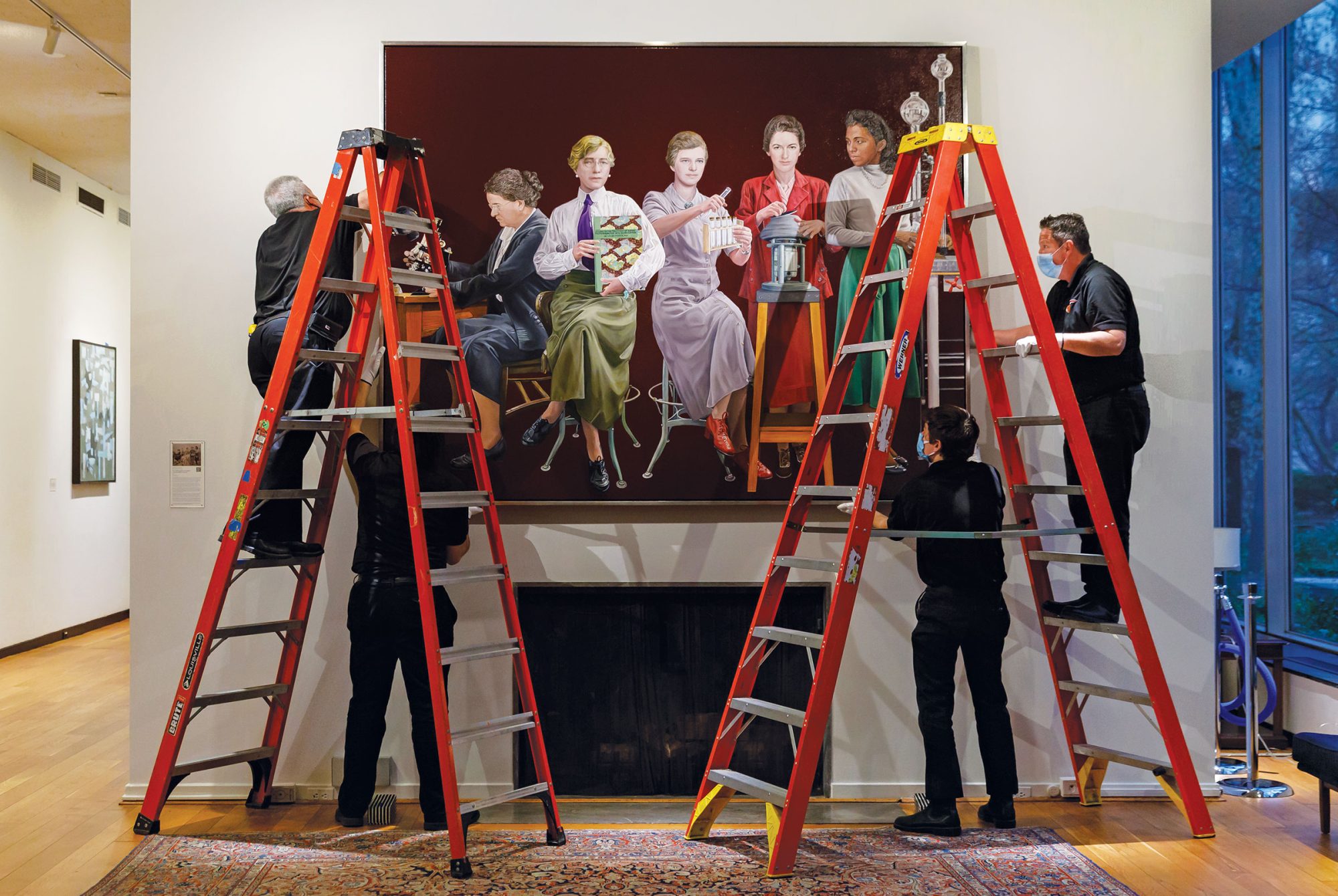
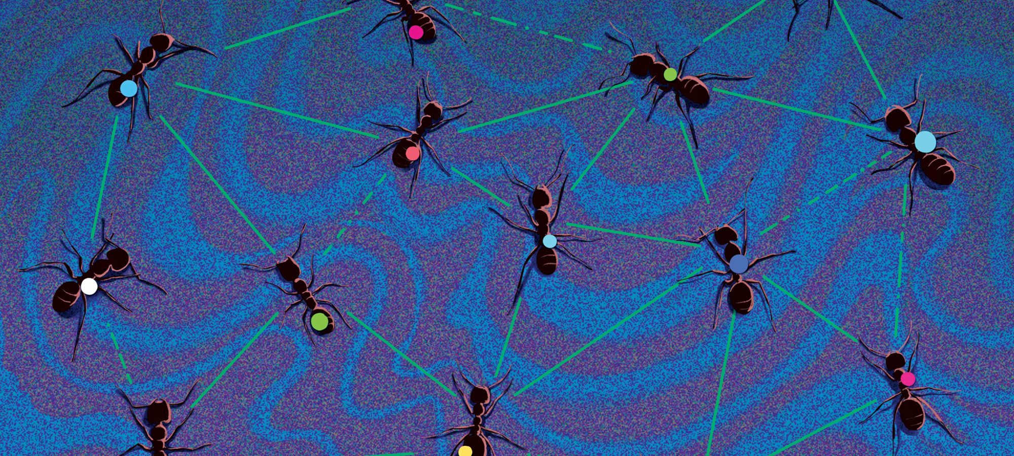
Single ants rarely make headlines, but ant colonies are capable of incredible things. Watch them transform a pile of dirt into an elaborate ant hotel, for example, and you might think that a single mind is steering the entire colony. In fact, this may not be far from the truth, according to recent work from Daniel Kronauer’s Laboratory of Social Evolution and Behavior.

The researchers found that ants can behave in much the same way that neurons collaborate in the brain, with regard to a process called sensory thresholding. Common to virtually all animals, sensory thresholding is a kind of cost-benefit analysis in which the organism reacts to a sensory input only when that stimulus crosses a certain threshold.

For instance, sensory thresholding may be at play when you decide to move out of a hot room. The point at which you’ll get up and leave will depend partly on the rising temperature and partly on internal factors, like the body’s need to preserve energy. You may initially stay put, but once the room gets hot enough to justify the hassle, you’ll head for the door.

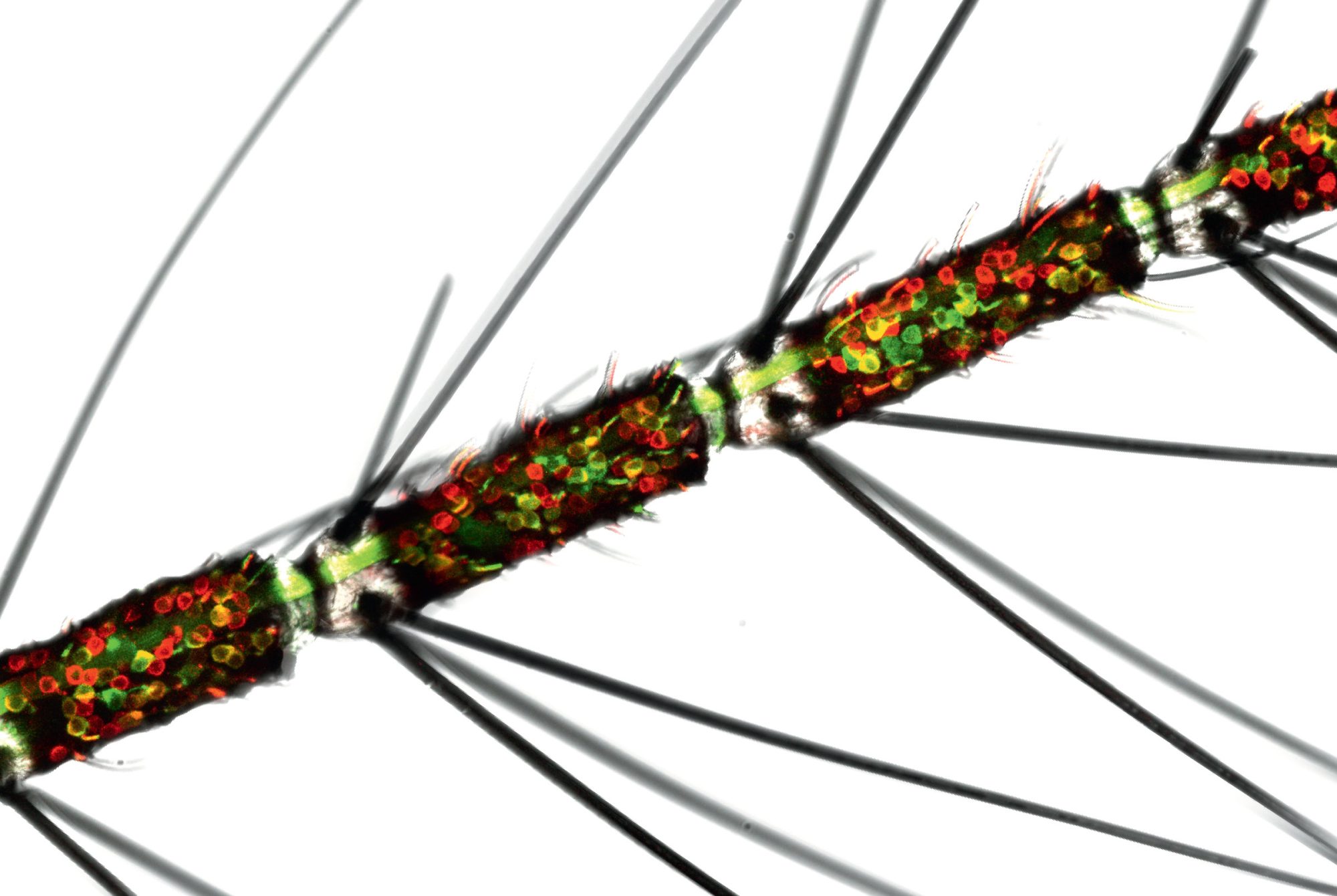
Kronauer and postdoctoral associate Asaf Gal wondered if ants would engage in sensory thresholding as a group, similar to the way neurons do in a brain—putting the needs of the whole network over that of individual cells. Working with clonal raider ants, Kronauer and Gal marked each ant with color-coded dots, let them form a nest, turned up the heat of the nest in precise increments, and tracked the ants’ responses.

Predictably, the ants fled the nest when temperatures reached uncomfortable levels, but they behaved more like a neural network than like humans shuffling out of a room. Specifically, how hot the nest had to become before the insects made an antline for the exit depended on the size of the colony: Those with around 50 members consistently fled at around 34 degrees Celsius, while colonies of 200 held out until the temperature reached 36 degrees.

This phenomenon is hard to explain if you think of ants as isolated individuals—an ant doesn’t know how many peers it lives with, so how can its decision depend on colony size? Kronauer and Gal suspect that pheromones, the messengers passing information between ants, scale their effect when more ants are present.

Still, why larger colonies require higher temperatures to pack up shop remains unclear. “It could simply be that the larger the colony, the more onerous it is to relocate, pushing up the critical temperature for which relocations happen,” ventures Kronauer, Rockefeller’s Stanley S. and Sydney R. Shuman Associate Professor.

The findings, reported in Proceedings of the National Academy of Sciences, suggest that ants combine sensory information with the parameters of their collective to arrive at a group response. And according to Kronauer, the research is “one of the first steps toward really understanding how insect societies engage in collective computation.”

A fuller picture
Photo by Will Ragozzino

On Campus

A fuller picture

Photo by Will Ragozzino

Thirty-five portraits line Rockefeller’s halls, most showcasing men scientists. The recently unveiled 36th tells a more inclusive story, highlighting five trailblazing women scientists. From left, there’s Florence Sabin, who advanced understanding of the immune system’s response to tuberculosis; Louise Pearce, creator of the first effective treatment for African sleeping sickness; Rebecca Lancefield, at the center, classified subtypes of strep bacteria, then ravaging WWI battlefields; next, Gertrude Perlmann deciphered pepsin’s 3D shape and its role in digestive disorders; Marie Maynard Daly, the first Black woman in the U.S. to receive a Ph.D. in chemistry, was instrumental in linking hypertension and high cholesterol to an increased risk of heart attack.

We need our fat
A blob of fat is as worthy of investigation as any other tissue. Jacob Pritchard

Forefront

We need our fat

A blob of fat is as worthy of investigation as any other tissue. Jacob Pritchard

There’s more to fat than meets the eye. We tend to think of our adipose tissue as being as unneeded as it is unwanted, nothing more than the much-maligned result of eating more calories than we burn. But scientists have come to realize that fat isn’t just a byproduct of metabolism; it’s also one of the key places where it happens. In fact, it would be better to think of fat tissue not as padding but as a complex, full body organ—with its own constellation of immune cells and nerve projections—that’s in constant dialogue with the endocrine system.

We need our fat, just like we need our intestines, liver, and stomach. And just like any organ, fat can suffer damage, with serious consequences.

Paul Cohen, a physician-scientist on staff at Memorial Sloan Kettering Cancer Center who studies obesity and its comorbidities, began to suspect that fat might be involved when survivors of childhood cancer showed up in his office with cardiometabolic diseases.

“I kept returning to this distinct group of patients,” he says. “They were developing coronary heart disease or diabetes at younger ages than expected in the absence of typical risk factors like obesity.”

Though Cohen’s patients presented as seemingly healthy young adults, all with normal BMIs and waist-to-hip ratios, they were already displaying the subtle indicators of brewing metabolic disease, such as rising blood sugar. Moreover, their fat tissue was brimming with immune cells and proteins known to be elevated in response to chronic injury.

And Cohen’s lab identified another commonality among these cancer survivors: As kids, they had all been treated with abdominal or total body irradiation. His hypothesis: Early exposure to radiation may cause long-term dysfunction in fat cells that manifests decades later.

He hopes that these findings, published recently in JCI Insight, will make clinicians rethink what they think they know about our metabolic systems. “When physicians are planning radiation therapy, they are very conscious of avoiding damage to major organs,” says Cohen, the Albert Resnick, M.D. Associate Professor at Rockefeller. “But fat is often not considered.”

A memorable walk in VR
Terceros at the VR station where mice create new memories. John Abbott

Forefront

A memorable walk in VR

Terceros at the VR station where mice create new memories. John Abbott

Neuroscientists have long known that memories are formed in the hippocampus, a small structure at the core of the brain. But recent findings suggest that’s only part of the story.

Researchers in the lab of Priya Rajasethupathy, the Jonathan M. Nelson Family Assistant Professor, found that a complex memory consists of a whole and its various details. You may bring to mind the full experience of last night’s dinner outing, for example, or just the taste of that ragù or a glimpse of candlelight. Working with mice, the scientists found that while the whole memory is stored right where they expected it—in the hippocampus—the fragments unexpectedly popped up in the prefrontal cortex.

To arrive at these discoveries, the researchers had to jump a few hurdles. Technical limitations have long hampered efforts to study memory as a distributed brain process. So Nakul Yadav, a graduate student in Rajasethupathy’s lab, built a novel setup using virtual reality to simultaneously record and manipulate neural activity from multiple brain areas. Perched atop a rolling Styrofoam ball, mice in the experiments strolled down an endless VR corridor, encountering various multisensory experiences along the way, each with its own pattern of lights, sounds, and smells. These sensory cues trained them to associate different “rooms” with pleasant or less-than-pleasant experiences. Nudged later by a specific sight or scent, the mice were able to recall the broader context and knew whether to happily expect sugar water or look out for an annoying puff of air.

In the process, the researchers discovered that a particular neural circuit makes two brain regions work in tandem during memory recall. This circuit lights up when prompted by the right kind of sensory input—a smell you come to associate with the meal, for example—and activates the prefrontal cortex, which then accesses the hippocampus for full memory retrieval. The work points to a new understanding of how the brain processes a memory.

“It suggests that there’s a dedicated pathway for memory recall, separate from memory formation,” says Andrea Terceros, a co-author of the study the team published in Nature. The findings might ultimately inform the treatment of dementias such as Alzheimer’s, which may be less about deficient memory storage than a breakdown in memory recall.

TB’s mutation mistake
CNRI

Forefront

TB’s mutation mistake

CNRI

In Southeast Asia, one particularly virulent tuberculosis strain infects half a million people each year. There may soon be a simple solution to that problem: macrolide antibiotics.

Number 1952

Data

The year when the original macrolide antibiotic, erythromycin, was first used. There are now three FDA-approved drugs of this class.

True, macrolides have historically failed to treat TB. But if the bacteria were to mutate in a particular way, TB would crumble in the face of these FDA-approved drugs—and in Southeast Asia, that’s exactly what appears to have happened. Jeremy M. Rock and colleagues serendipitously discovered that the Southeast Asian strain picked up precisely the right mutation about 900 years ago—rendering it vulnerable, in theory, to a class of readily available drugs. Since publishing findings in Nature Microbiology, Rock, who is Rockefeller’s Penrhyn E. Cook Assistant Professor, has been devising a way to apply them in a clinical setting, an effort that could ultimately save thousands of lives.

Math on the fly
Vector math is integral to the brain’s navigational functioning. Illustration by Israel G. Vargas

Forefront

Math on the fly

Vector math is integral to the brain’s navigational functioning. Illustration by Israel G. Vargas

You’re walking down the street on a sunny autumn day. You hear a bang and, without skipping a beat, turn your head to see what’s going on: Someone slammed a car door. Sound identified, you march on.

We usually take for granted our ability to walk in one direction while facing another without getting disoriented. But perhaps we shouldn’t­; in the lab of Rockefeller’s Gaby Maimon, scientists are fascinated by the brain’s ability to construct a sense of spatial orientation as we move through the world.

Recently, Maimon and graduate student Cheng Lyu discovered a set of math-savvy neurons in fruit flies that might reveal how the animals keep heading in the right direction. These neurons, of which there are four classes, perform complex mathematical calculations along four axes to indicate the fly’s traveling direction. In work reported in Nature, the researchers found that each neuronal class can be thought of as representing a mathematical vector whose angle points in the direction of its associated axis. A vector’s length indicates how fast the fly is moving in that direction.

“Amazingly, a neural circuit in the fly brain rotates these four vectors so that they are aligned properly to the angle of the sun and then adds them up,” Maimon says. “Neuronal circuits implement relatively sophisticated mathematical operations.”

These cells light up when you’re down for the count
These neurons provide clues to what the brain is up to when the body fights a pathogen. Laboratory of Molecular Genetics

Forefront

These cells light up when you’re down for the count

These neurons provide clues to what the brain is up to when the body fights a pathogen. Laboratory of Molecular Genetics

It’s easy to relate to a mouse feeling under the weather. It seeks out a safe spot to hide, preferring not to move, and it doesn’t have much of an appetite.

In fact, most animals will avoid moving, eating, and drinking when they’re fighting an infection. It’s the smart thing to do—these near-universal sickness responses allow the organism to save energy that the immune system needs to fight off the pathogen. But precisely how the behaviors are orchestrated was an open question when researchers in the lab of Jeffrey M. Friedman, the Marilyn M. Simpson Professor, set out to search for neural activity induced by an immune response.

A cluster of neurons lit up in the brain stem whenever the scientists provoked a mouse’s immunity. When firing, these cells subdued the animal’s movement, eating, and drinking; and when the researchers directly triggered the same neurons in healthy mice, those animals began displaying similar behaviors. The findings were published in Nature in October.

Anoj Ilanges, a former graduate student in Friedman’s lab who is now a group leader at the Howard Hughes Medical Institute’s Janelia Research Campus, says that little is known about the central nervous system’s role in infection. “We looked at one region of the brain,” he says, “but there are many others that become activated with the immune response. This opens the door to asking what the brain is doing holistically during sickness.”

New blood in the copycat club
ISTOCK / LEON GIN

Forefront

New blood in the copycat club

ISTOCK / LEON GIN

PRECIOUS FEW ANIMALS can learn to imitate new sounds, a skill known as advanced vocal learning. Humans and parrots can do it, as can whales, seals, bats, hummingbirds, songbirds, and elephants, the last of which have been observed copying the sounds of passing trucks. New research from the laboratory of Erich D. Jarvis welcomes woodpeckers as members of this rather exclusive club, albeit for a different reason.

Jarvis and his team were surprised to discover that woodpeckers possess specialized neural circuits that resemble the brain structures that allow young songbirds to learn new tunes. However, the woodpecker brain regions were activated not by vocalization but by drumming on tree trunks—a rhythmic behavior that the birds use to compete for territory. Writing in PLOS Biology, the team proposed that the woodpecker drumming circuit and the vocal learning pathways of songbirds and humans evolved from the same ancestral structure.

All vaccines on deck
Pfizer, Moderna, Johnson & Johnson, AstraZeneca, Gamaleya, CanSino, or Sinovac? ISTOCK / MARSBARS

Forefront

All vaccines on deck

Pfizer, Moderna, Johnson & Johnson, AstraZeneca, Gamaleya, CanSino, or Sinovac? ISTOCK / MARSBARS

“Did you get Pfizer or Moderna?”

Here in the United States, this question became something of an icebreaker. But in Mexico, where officials administered seven different COVID vaccines, it would not have rolled off the tongue quite so easily: Pfizer, Moderna, Johnson & Johnson, AstraZeneca, Gamaleya, CanSino, or Sinovac?

“Mexico allowed concomitant use of different vaccines, including those not yet approved by the World Health Organization, since there was insufficient vaccine production to meet the demand,” says Santiago Avila-Rios, an assistant professor at the National Institute of Respiratory Diseases in Mexico.

To what extent this motley approach to population immunity worked had been unknown, however, since the vast majority of research on vaccine efficacy has been done on the big-name mRNA vaccines, Pfizer and Moderna. So Rockefeller virologists Theodora Hatziioannou and Paul Bieniasz teamed up with Avila-Rios and other researchers in Mexico to find out.

Number 198

Data

Number of doses of COVID vaccine administered per 100 people in the United States. In Mexico, that number is 174; in Cuba, it’s 335.

“They were using many vaccines that we haven’t seen in the U.S.,” Hatziioannou says.

There were clear concerns that the lesser-known vaccines would offer inferior protection compared with big names like Pfizer and Moderna, yet the researchers didn’t discount the possibility that some of those unsung vaccines might actually work better. “Some vaccines involved different methods of inducing immunity—adenovirus vectors, whole activated viruses, multiple doses,” Hatziioannou says. “So we wanted to know what level of neutralizing antibodies were achieved with these other methods.”

As it turned out, the vaccines elicited a range of immune responses: Pfizer was the strongest; Sinovac, the weakest. None provided much neutralizing activity against the omicron variant in patients who had never been exposed to the virus, but all the vaccines produced neutralizing antibodies against omicron in those who had been infected either before or after vaccination.

The results were heartening at the population level, where the data suggests that low- and middle-income countries will be able to achieve immunity by hook or by crook—by vaccinating and boosting individuals with whichever vaccines are available, or through repeated exposure via infection. “Even in the absence of the best vaccines, it might be possible to achieve population immunity with a mix of approaches,” Hatziioannou says about the study, published last year in the journal mBio. “We expect that low- and middle-income countries will ultimately achieve good levels of immunity against the virus, though they might need to prioritize boosters for people who received vaccines that elicit low levels of antibodies.”

The benefit of boosters
ISTOCK

Forefront

The benefit of boosters

ISTOCK

Omicron variants have been stalking the globe for over a year, and they’re wildly infectious. If you’re overdue for a booster, now’s the time to get one. A team led by Paul Bieniasz, Theodora Hatziioannou, and Michel C. Nussenzweig reviewed blood samples from individuals who had received second and third doses of an mRNA vaccine, and they found up to 200-fold increases in neutralizing activity against the omicron variant. They reported their results in the New England Journal of Medicine.

“Our study makes it clear why the third dose should be recommended,” says Nussenzweig, the Zanvil A. Cohn and Ralph M. Steinman Professor. “It’s one of the best reactions to the virus that we’ve seen.”

How misfolded proteins get into shape
Illustration by Matt Chase

Forefront

How misfolded proteins get into shape

Illustration by Matt Chase

The proteins in our bodies are constantly twisting themselves into the most exquisite origami. Flipping and folding just so, strings of amino acids take on precisely the right forms—pleats, horseshoes, jelly rolls—to keep our functions beautifully humming along. Except when they don’t—and one misshapen molecule can spell catastrophic system failure.

Cystic fibrosis is a prime example. Doctors have long understood exactly how the disease does its devastating work: At its heart is CFTR, a protein channel lying atop cells lining the lungs and digestive tract that attracts water to thin and move mucus. But if CFTR doesn’t fold correctly, it can barely function at all. Then mucus accumulates and hardens, breathing and digestion become painfully difficult, and the lungs become a fertile ground for pathogens.

DNA Illo

Data

Approximately 2,500 mutations in the CFTR gene have been linked to cystic fibrosis.

Fortunately, powerful drugs called correctors can significantly prolong patients’ lives. Until recently, no one truly understood how they worked—until Jue Chen, Rockefeller’s William E. Ford Professor, and her team demonstrated how one medication known as a CFTR corrector directly addresses the misfolding.

They did it by stitching together thousands of snapshots of the corrector in action, developing a clear picture of how the drugs stabilize CFTR by nestling into a notch within the protein. That understanding enabled them to develop a theory, published last year in Cell and Science, of how protein folding correctors do their job. As it turns out, Chen’s group may have opened the door to treatments for a range of heretofore intractable conditions.

Hundreds of diseases, from Parkinson’s to sickle cell anemia, occur when proteins fail to assume the correct 3D structure. “We now have a way to identify molecules that may be used to treat these diseases,” Chen says.

Two diseases, one problem
At the molecular level, tumors of heavy smokers look a lot like those of patients with Fanconi anemia, pictured. Laboratory of Genome Maintenance

Forefront

Two diseases, one problem

At the molecular level, tumors of heavy smokers look a lot like those of patients with Fanconi anemia, pictured. Laboratory of Genome Maintenance

SCIENTISTS HAVE FOUND an intriguing relationship between patients with Fanconi anemia, a rare genetic disorder, and people without the disease who smoke cigarettes: Both are vulnerable to a certain kind of genetic havoc, increasing their risk for developing head and neck squamous cell carcinoma, a common cancer growing in the mucous membranes of the mouth, nose, and throat.

In work published in Nature in November, the lab of Agata Smogorzewska found that Fanconi patients’ cells are unable to repair DNA damage caused by chemicals called aldehydes found in some foods and in cigarettes. As a result, their genomes acquire structural problems—a sort of Goldilocks syndrome in which genes are present in too many or too few copies, with stretches of DNA appearing in the wrong places or not at all, creating a perfect storm for the development of quickly metastasizing tumors.

As it turns out, similar genomic defects are observed in tumors from people without Fanconi anemia, and the researchers found a correlation between these individuals’ smoking history and the frequency of structural variants: The more a person had smoked, the more variants were detected in their tumors. Smogorzewska posits that smoking subjects the body to so much aldehyde-induced damage that otherwise healthy repair mechanisms fail to keep up.

“So, cells from people without Fanconi anemia act as if they too have a DNA-repair defect,” she says. “This rare disorder may be telling us something profound about how certain cancers are triggered in the general population.”

A scent paradise for flies
Photograph by Matthew Septimus

Gadget

A scent paradise for flies

Photograph by Matthew Septimus

nothing gets Drosophilae going like the tangy perfume of apple cider vinegar, which evokes the scent of rotting fruit. From atop a spinning ball at the center of a virtual reality setup, the insect will run or fly toward the scent while Chad Morton and Andrew Siliciano, both graduate students in Vanessa Ruta’s’ Laboratory of Neurophysiology and Behavior, observe the neural activity enabling it to navigate an aroma-rich world.

To craft this “odorverse,” the scientists retrofitted a fly treadmill system developed in the lab of Gaby Maimon with a 3D-printed plate that allows the fly to rotate as it walks about its fictive environment. Working closely with engineers in the Precision Instrumentation Technologies shop, they then added a pistol-shaped nozzle for odor release that connects to airflow-modulating controllers and scent vials. “We used Python to program the software that manipulates the odor dispenser,” says Morton. “That way,” adds Siliciano, “we can precisely measure the amount and vary the concentrations of the odor we’re releasing.” (In between experiments, they might expel a puff of 1-octanol acid, a palate-cleansing neutral fragrance.)

Until recently, the only way scientists could manipulate individual scents was to turn them on or off. But the high-precision scent diffuser makes it possible to more accurately simulate how a fly might navigate amid multiple smells, so scientists can explore what happens when it loses a scent trail and the decisions it makes along the way.

These experiments are already revealing that a fly’s path to an aroma is more elaborate than previously thought. Rather than tracking straight up the center of a plume, it prefers to wiggle in and out of the plume’s periphery, perhaps taking a whiff of surrounding scents to stay open to other environmental inputs.

And while the fly’s goal may be to land on a juicy grape, Morton and Siliciano are after something more elusive: the circuits that fire as the fly smells its way around—intel that might tell us more about the basic functioning of sensory systems.

Highly receptive
Laboratory of Neurogenetics and Behavior

Snapshot

Highly receptive

Laboratory of Neurogenetics and Behavior

The neuroscience of smell is supposed to be straightforward. Each olfactory neuron expresses a single olfactory receptor unique to a specific kind of scent. This streamlined system shows up across a variety of species, from flies to mice to humans.

But while studying Aedes aegypti mosquitoes, Leslie B. Vosshall and colleagues discovered that these insects aren’t playing by the rules—and, as a result, have evolved a uniquely resilient sense of smell. This close-up of a mosquito antenna reveals an olfactory system that runs counter to conventional wisdom: Some odor neurons (shown in red and green) express one receptor, while others (yellow) are going rogue, displaying multiple receptors. “This was very surprising,” says Vosshall, the Robin Chemers Neustein Professor, about her findings. “Mosquitoes unexpectedly pack a very large number of receptors into a single smell neuron.”

Vosshall notes that it’s very hard to tamper with such a complex biological system, which may explain why she and others in the field have long struggled to come up with ways to manipulate a mosquito’s sense of smell. Conceivably, a better understanding of how the system works could open the door to developing effective repellents or other tools to stop Aedes and other such pests from homing in on humans.