Learning to see the world differently
Illustration by Angie Wang

Forefront

Learning to see the world differently

Illustration by Angie Wang

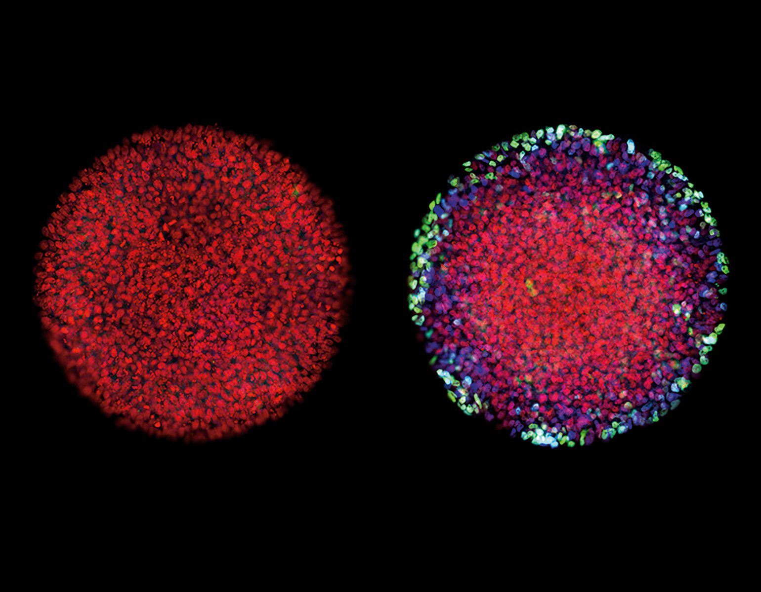
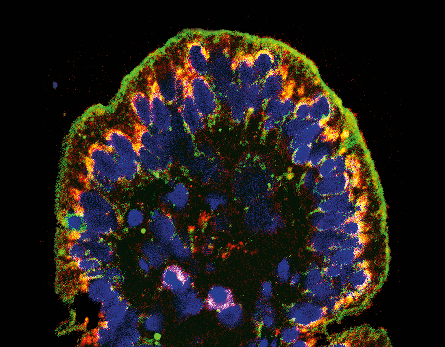
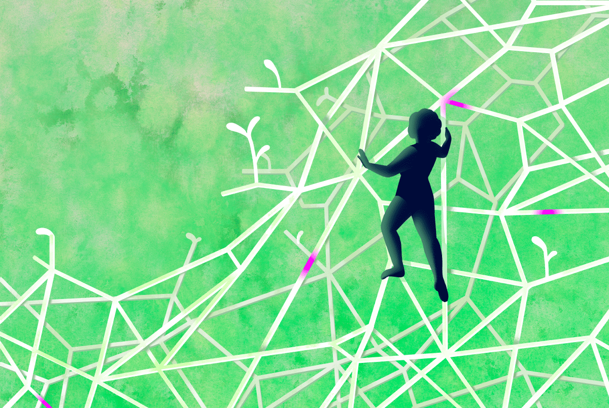
Personal growth begets neuron growth. As you learn a language or learn to hang glide, for instance, brain cells sprout new appendages, known as axons, that send signals to other cells. Researchers have long been aware of the brain’s capacity to reconfigure itself, but it is less clear how this quality, known as plasticity, supports various aspects of learning.

Learning to see - plant illo

Data

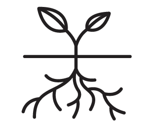
Animals have some plasticity; plants have a lot. Being able to change in response to your environment is especially beneficial if you’re rooted to the ground, hence unable to escape it.

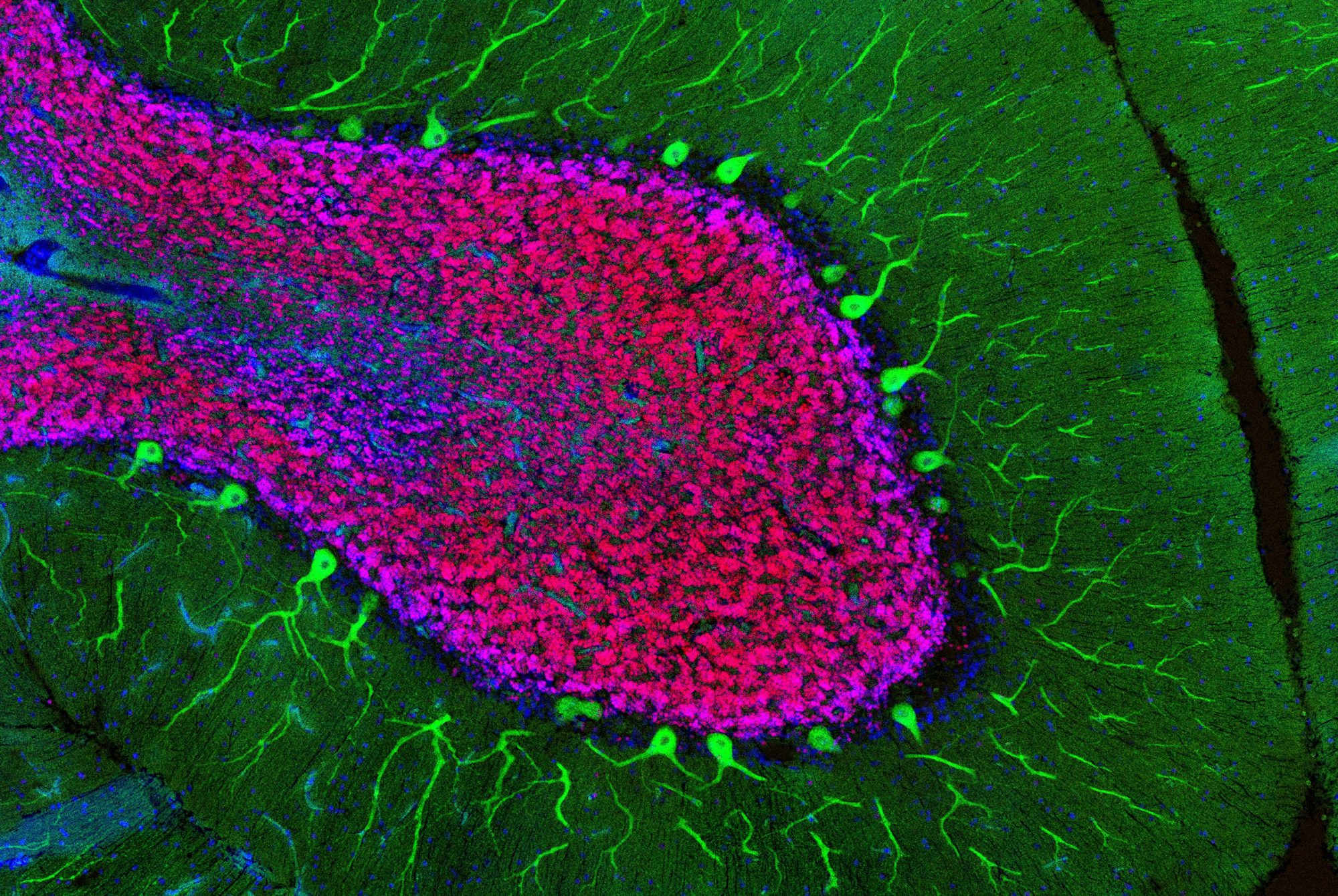
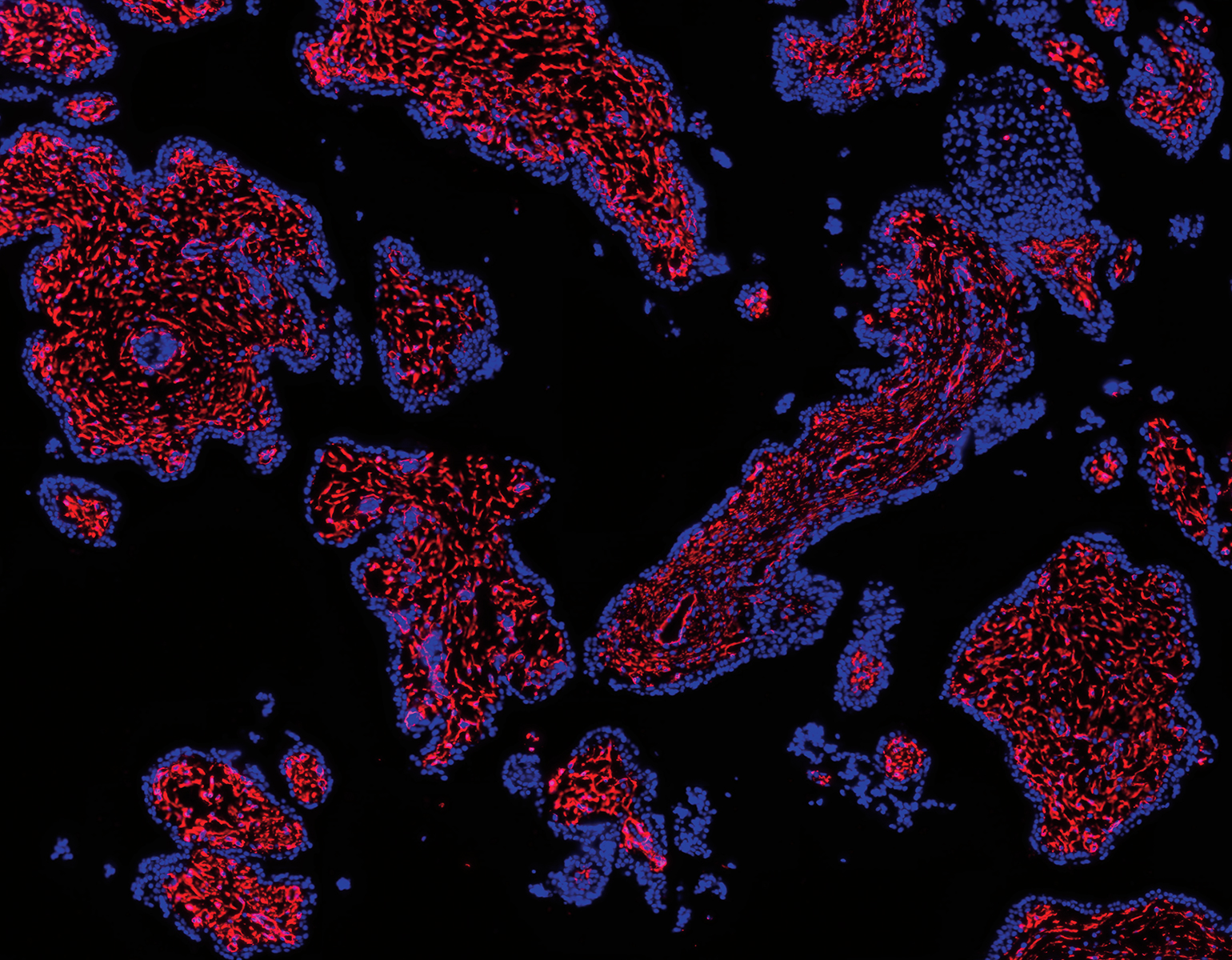
In teaching macaque monkeys new visual skills, Charles D. Gilbert and his colleagues were able to study how axons grow during perceptual learning, a process that tunes the brain to more adeptly detect certain sights, smells, or sounds. The researchers showed the monkeys busy patterns within which, with a trained eye, lines could be traced. As the monkeys got better at spotting the lines, the researchers found, their neurons grew fresh axons in the visual cortex, a brain area that processes signals received from the eye. This experiment, described in Proceedings of the National Academy of Sciences, offers a fresh look at the precise manner in which experiences change how the brain perceives and responds to the environment.

“We’ve always known the brain needs some degree of plasticity through adulthood,” says Gilbert, the Arthur and Janet Ross Professor, “but it turns out that plasticity is more widespread than we initially thought.”

Brain botany
The Rockefeller University / Laboratory of Molecular Biology

Snapshot

Brain botany

The Rockefeller University / Laboratory of Molecular Biology

There are billions of cells in the human brain whose special features scientists are just beginning to understand. In other words, it’s a jungle in there. Xiao Xu, Elitsa Stoyanova, and Maria Moya obtained this image of the human cerebellar cortex with antibodies marking two species of brain cell: Purkinje cells (green) and granule cells (red). The three are graduate fellows in the lab of Nathaniel Heintz and are working on a new method to isolate specific classes of neurons, especially those related to neurodegenerative disease.

More than one way to kill a microbe
A new drug kills antibiotic-resistant bacteria by destroying their cell walls (green). Photo by Dr. Kari Lounatmaa / Science Photo Library

Forefront

More than one way to kill a microbe

A new drug kills antibiotic-resistant bacteria by destroying their cell walls (green). Photo by Dr. Kari Lounatmaa / Science Photo Library

It’s not just humans that kill bacteria. For bacteriophages, a type of virus, microbe murder is central to survival. In nature, these viruses invade bacteria, replicate inside of them, and then liberate their progeny through the release of lysins, enzymes that dissolve the bacteria’s cell walls.

Vincent A. Fischetti has spent the past 20 years studying the bacteria-bursting properties of lysins. His work has long yielded promising results in animal experiments, and now an early clinical trial suggests that this type of treatment could also work in humans.

Sponsored by the biotech company ContraFect, the phase II trial involved patients with methicillin-resistant Staphylococcus aureus, or MRSA, a common hospital infection that doesn’t respond to conventional antibiotics. The researchers found that, among people whose infection had spread to the blood, the response rate to treatment was 40 percent higher when a lysin-based drug called exebacase was given together with antibiotics, compared to when antibiotics were administered alone.

These findings bring new hope to researchers and clinicians seeking a different way to combat bacterial infections. “Bacteria are growing more and more resistant to antibiotics,” says Fischetti, “and we’re showing that there are other ways to fight them.”

When cancer cells cut corners, it can be their downfall
The Rockefeller University / Laboratory of Metabolic Regulation and Genetics

Forefront

When cancer cells cut corners, it can be their downfall

The Rockefeller University / Laboratory of Metabolic Regulation and Genetics

Cancer cells are, by definition, abnormal. But some are odd even by cancer’s standards. There are, for instance, those that fail to produce vital nutrients.

Cells that cause a rare form of lymphoma, called ALK-positive ALCL, have forfeited the ability to make their own cholesterol in order to focus on more grandiose tasks, such as wreaking havoc on the body. They compensate for their metabolic deficiency by stealing nutrients from the surrounding environment. For Kivanç Birsoy, the Chapman Perelman Assistant Professor, it’s a vulnerability that might offer an alternative way to treat the disease, which can grow resistant to chemotherapy.

When cancer cells cut corners, it can be their downfall

Data

The fraction of a cell's outer membrane that is made of cholesterol. The substance keeps the membrane durable without being rigid, allowing the cell to move and flex without breaking.

In research reported in Nature, Birsoy’s team created a line of ALCL cells lacking receptors for cholesterol uptake to see how they would cope without access to the nutrient. The cells died almost immediately.

“We think therapies that block uptake of cholesterol might be particularly effective against chemotherapy-resistant forms of ALCL,” says Birsoy, “and they might be useful for some other cancer types as well.”

Embryo cells live and learn
Embryo cells on the right are unable to diversify because they haven't received the right signals. Laboratory of Theoretical Condensed Matter Physics

Forefront

Embryo cells live and learn

Embryo cells on the right are unable to diversify because they haven't received the right signals. Laboratory of Theoretical Condensed Matter Physics

New research suggests that we are—quite literally—shaped by our earliest life experiences. For the past 25 years, scientists have believed that when cells in the embryo begin to specialize into gut cells, brain cells, or other cell types, they are obeying the instruction of a single signaling protein called activin. Recently, however, graduate fellow Anna Yoney realized things are not quite this simple.

In a new study, published in eLife, Yoney, along with Eric D. Siggia and Ali H. Brivanlou, found that activin does set off the specialization process, known as differentiation, but only in embryo cells with particular past experiences. Working with artificial human embryos, the researchers found that cells differentiated only if they were exposed to a different chemical, WNT, before being exposed to activin—a phenomenon the researchers termed “signaling memory.”

Embryo cells live and learn

Data

The time it usually takes for a fertilized human egg to become a clump of differentiating cells, called blastocyst.

Until now, scientists failed to notice the role of WNT because, says Yoney, most developmental biologists work with animal cells.

“Scientists had been watching activin induce differentiation for decades—in mouse cells, frog cells, and in other model organisms,” Yoney says. “But the problem with animal cells is that they’ve already encountered a number of cellular signals. Our artificial embryos hadn’t had that kind of exposure.” (Read more about embryo research in “Science, society, right and wrong”.)

The aquatic superpowers of geckos
To speed across ponds without sinking, geckos rely on buoyancy, movement, and the ability of their skin to repel water. Photo by Pauline Jennings

Forefront

The aquatic superpowers of geckos

To speed across ponds without sinking, geckos rely on buoyancy, movement, and the ability of their skin to repel water. Photo by Pauline Jennings

Geckos shouldn’t be able to walk on water. Outside the realm of biblical miracles, water walking is typically reserved for two types of animals: those small enough to balance on water’s surface tension, and those large enough to hoist themselves above the water through sheer force. A comfortably midsize animal, the gecko doesn’t fall into either of these categories.

And yet Jasmine Nirody found herself watching a video of a gecko that seemed to be easily traipsing across water. A Rockefeller fellow in physics and biology, she immediately began investigating this spectacle.

Swimming is a great way to get around—unless someone is chasing you. For those situations, some water animals employ special techniques to skedaddle.

Working with colleagues at the University of California, Berkeley, Nirody found that, like bigger lizards, geckos use a slapping motion to pull their bodies above water. And, like spiders, they take advantage of water’s surface tension. In other words, they combine techniques from opposite ends of the size spectrum to stay afloat. Further, the scientists discovered that geckos have a feature all of their own that contributes to their aquatic agility.

“Geckos have this amazing superhydrophobic skin that repels water and enhances their ability to stay above the surface,” says Nirody.

In addition to elevating our respect for reptiles, this research could be used to create tools with real-world applications. “Our work with animal locomotion is geared toward use in robotics,” says Nirody. “And an intermediate-sized water-running robot, for example, would be ideal for searching flooded areas after a natural disaster.” 

Online recipes for classroom science
Photo by Mario Morgado

Forefront

Online recipes for classroom science

Photo by Mario Morgado

At bard high school Early College Queens science educator Stephanie Kadison (center) is always seeking fresh ways to engage her students. Along with other New York City teachers, Kadison recently collaborated with Rockefeller’s Science Outreach program to help develop a new online resource available to learners, educators, and scientists everywhere.

The website, RockEDU Online, features a versatile portfolio of science education materials supporting both teachers looking to enhance their classroom routine and scientists who want to engage with schools in their community.

Find it on rockedu.rockefeller.edu

Cellular “trafficker” linked to autism
Mary E. Hatten. Photo by Mario Morgado

Forefront

Cellular “trafficker” linked to autism

Mary E. Hatten. Photo by Mario Morgado

Things can get a bit hectic at synapses, the junctions where neurons connect. To manage an onslaught of incoming chemical signals, nerve cells must perpetually remove old receptors from their surface to make room for new ones, a process facilitated by molecules called protein traffickers.

When these proteins fail to do their job, the ensuing synaptic mess may negatively impact the brain’s development. Recently, Rockefeller’s Mary E. Hatten, the Frederick P. Rose Professor, and collaborators at Johns Hopkins University were able to illuminate the process by which defects in a trafficker known as ASTN2 may lead to autism and other neurodevelopmental conditions.

When studying the cerebellum region of mouse brains, the researchers found reasons to suspect that low levels of the protein might lead to weak neural connectivity and atypical brain function. Supporting this notion, the scientists identified a family in which three children carried ASTN2 mutations and additionally suffered from neurodevelopmental issues including autism and language delays. These findings, published in Proceedings of the National Academy of Sciences, are consistent with recent data from population studies linking ASTN2 mutations to a variety of brain disorders.

How to patch up an ailing intestine (quite literally)
The Rockefeller University / Laboratory of RNA Molecular Biology

Forefront

How to patch up an ailing intestine (quite literally)

The Rockefeller University / Laboratory of RNA Molecular Biology

People with Crohn’s disease and ulcerative colitis have a lot in common. For starters, they share a range of symptoms, such as stomach pain, diarrhea, vomiting, and weight loss. Their treatment tends to involve taking anti-inflammatory drugs. And they have similar reasons to be rather unhappy with those anti-inflammatories: the drugs often don’t work very well and come with unpleasant side effects.

Now there is hope for a better treatment, based on yet another common denominator of the two diseases: intestinal leakiness. Both Crohn’s disease and ulcerative colitis stem from weakness in a thin cell layer that lines the intestine. When this lining becomes porous, bacteria seep into the surrounding tissues and bowel inflammation ensues.

Research associate W. Vallen Graham thought there might be a way to fix this underlying plumbing issue; and in a search for compounds that block MLCK, a protein believed to undermine intestinal-wall tautness, he recently discovered one that does the trick. Results from experiments with mice, reported in Nature Medicine, may point the way for future therapies that boost the effectiveness of anti-inflammatories by sealing intestinal leaks.

“This is exciting, because there’s currently no drug that can remedy permeability of the intestine,” says Graham, who began the research at Harvard Medical School and is continuing it in the lab of Rockefeller’s Thomas P. Sakmar.

When breakthroughs break off

Forefront

When breakthroughs break off

the world has nearly forgotten Israel Kleiner. A late Rockefeller scientist active in the 1910s, Kleiner conducted pioneering research on diabetes and came close to discovering a lifesaving treatment. Close, but not close enough to bring his work to fruition or make a name for himself. World War I, and Kleiner’s bosses, interfered with his hopes of finding a cure for the mysterious disease. At the time, diabetes was claiming thousands of lives, but the university’s priorities lay elsewhere—on infectious diseases rampant among soldiers, for example.

It was only a decade later that other scientists, by building on Kleiner’s work, were able to show that the hormone insulin could be used to lower patients’ blood sugar levels. Kleiner, though, was out of luck, out of funding, and, eventually, out of a job.

Why should we remember the efforts of an obscure, century-old scientist? Because, argues Jeffrey M. Friedman, who chronicles Kleiner’s destiny in his article “Discovery, Interrupted” in Harper’s Magazine, the story of the late scientist’s exile holds important lessons for today’s society, where the value of open-ended research is being similarly challenged.

“Focusing too much on mainstream notions of what is important or useful carries the risk that the very discoveries that make translational research possible will never be made,” writes Friedman, the Marilyn M. Simpson Professor. “It also presupposes that we know what will be important in the future,” an idea for which little evidence exists.

Research on TB returns to earth
Photo by Jacob Pritchard

Forefront

Research on TB returns to earth

Photo by Jacob Pritchard

Half a century ago, when the antibiotic rifamycin was discovered in soil from a French pine forest, it led to the most potent treatment for tuberculosis ever developed. Unfortunately, victory didn’t last.

In recent years, the disease has made a crushing, antibiotic-resistant comeback. Amid scientists’ scramble to develop alternative treatments, TB’s fast-evolving pathogens are claiming millions of lives.

In looking for solutions, a team of Rockefeller scientists have gone back to the source from which rifamycin first emerged: Mother Nature.

“Rifamycin is naturally produced by a soil bacterium,” says Sean F. Brady, the Evnin Professor, who led the work. “So we wanted to find out whether nature had also made analogs of the compound—molecules that look like rifamycin, but that have slight differences.”

Sequencing the genes of microbes found in soil, Brady’s lab identified a group of natural antibiotics, known as kanglemycins, or kangs, that are closely related to rifamycin. Further analysis revealed that these antibiotics have structural features that set them apart from their cousin, including an extra sugar and an extra acid.

These tiny differences allow kangs to effectively combat mycobacteria that don’t respond to rifamycin. “We’d still like to see increased potency and broader activity against resistant bugs,” says research associate professor Elizabeth Campbell, who was also involved in the study, “but our findings tell us that we’re on the right track.” (Learn more in “TB is changing.”)

The new River Campus, unveiled
  1. The Bass Dining Commons, a café with indoor and outdoor seating and sweeping East River views, replaces the university’s 1971 lunchroom.

  2. Two laboratory floors are each 750 feet long. Horizontally-oriented lab buildings are good for science because they help spur informal collaboration: When people work on the same floor, they are more likely to work together.

  3. A huge landscaped roof brings the Rockefeller campus all the way to the water. The outdoor space also features a two-level amphitheater carved out of the western façade.

  4. At the water’s edge, the public East River Esplanade has been repaired and refurbished, with new pavers, benches, lights, and landscaping. Vehicle traffic flows by behind the sound barrier.

  5. The Hess Academic Center provides spacious new executive offices and two mid-sized conference rooms.

  6. A conference facility for up to 100 guests, the Kellen BioLink, hosts retreats and small symposia. Sliding glass walls open onto the Fascitelli Great Lawn.

On Campus

The new River Campus, unveiled

  1. The Bass Dining Commons, a café with indoor and outdoor seating and sweeping East River views, replaces the university’s 1971 lunchroom.

  2. Two laboratory floors are each 750 feet long. Horizontally-oriented lab buildings are good for science because they help spur informal collaboration: When people work on the same floor, they are more likely to work together.

  3. A huge landscaped roof brings the Rockefeller campus all the way to the water. The outdoor space also features a two-level amphitheater carved out of the western façade.

  4. At the water’s edge, the public East River Esplanade has been repaired and refurbished, with new pavers, benches, lights, and landscaping. Vehicle traffic flows by behind the sound barrier.

  5. The Hess Academic Center provides spacious new executive offices and two mid-sized conference rooms.

  6. A conference facility for up to 100 guests, the Kellen BioLink, hosts retreats and small symposia. Sliding glass walls open onto the Fascitelli Great Lawn.

the views up and down the East River are inspiring. The breeze, slightly salty, is a pleasure. But standing atop Rockefeller’s new campus extension, the most remarkable thing is what you don’t see, don’t hear, and don’t smell: a six-lane urban highway choked with over 100,000 vehicles a day. It’s gone without a trace, expertly buried underneath two acres of landscaped green space.

The disappearance of the roadway that has formed Rockefeller’s eastern border since the 1940s is just one of the benefits of building over the FDR Drive. More importantly, by siting new construction in the Drive’s unused air rights, it is possible to construct a lab building with a unique shape—long and low—that would not otherwise be possible in a dense urban environment.

This is the Stavros Niarchos Foundation–David Rockefeller River Campus, a two-acre parcel of artificial land, and the Marie-Josée and Henry R. Kravis Research Building, a lab building suspended in midair. Nearly four blocks long, the Kravis Building is just two stories high, making it well-suited to the need of modern collaborative science. It’s the new home of 18 Rockefeller labs, with space for five more.

The building’s landscaped rooftop— accessible both from within the Kravis Building and from the existing campus via two sets of low-slung exterior stairs—is the center of the expanded campus. With ample riverside seating and pleasant landscaping, it’s an amenity in and of itself.

“This project is transformational,” says Richard P. Lifton, Rockefeller’s president. “It is yielding spectacular laboratory space that will house a third of our faculty, a rooftop dining hall, administrative building, and gardens, that all provide beautiful vistas overlooking the East River. The next generation of great scientists will make their key discoveries here.”

And underneath it all, the city traffic crawls imperceptibly along.

Photographs by Halkin Mason Photography
A meticulous map of the human placenta
Stromal and endothelial cells of the plancenta, in red. Laboratory of RNA Molecular Biology, The Rockefeller University

Forefront

A meticulous map of the human placenta

Stromal and endothelial cells of the plancenta, in red. Laboratory of RNA Molecular Biology, The Rockefeller University

Few of the body’s organs are as hard at work as the placenta during its first three months of service: It feeds and protects the fetus while supplying antibodies, hormones, and blood. Yet little is known about this earliest chapter in the mother-child relationship. Now, researchers have developed a new way to analyze the microscopic interactions between fetal and maternal cells.

A meticulous map of the human placenta

Data

At full-term pregnancy, the placenta filters up to three cups of blood per minute.

Thomas Tuschl and his colleagues recently performed an in-depth survey of human placental and decidual tissues, which contain cells from the fetus and mother, respectively. The researchers identified 20 distinct cell types, and, using a unique RNA-sequencing strategy, made inventories of genes associated with each type.

The end result, according to postdoc Hemant Suryawanshi, is the first “cellular atlas” of the early human placenta—a map that, among other things, will help scientists pinpoint causes of pregnancy complications.

“In pregnancy, there are dramatic changes both in cellular composition and at the molecular level,” says Suryawanshi, who together with his colleagues reported these findings in Science Advances. “Now, for the first time, we have high-resolution pictures of those changes.”

The Sharpie
Video by Daniel Kronauer

Gadget

The Sharpie

Video by Daniel Kronauer

without the sharpie, science would grind to a halt. Samples would get mixed up, data would go unrecorded, glassware would disappear into neighboring labs. A staple of the laboratory since the 1960s, the Sharpie is prized for its ability to reliably write on just about anything. Its utility makes it ubiquitous: Rockefeller scientists alone go through some 6,500 Sharpies a year.

Sharpie pen illustration

In neuroscientist Daniel Kronauer’s lab, they have even become experimental equipment. It turns out that Ooceraea biroi ants, the species used in much of Kronauer’s work, shun fresh Sharpie ink. That makes Sharpies a great tool for confining ants to small areas, or for testing their olfactory function. (For more on Kronauer’s research, see “Even small brains make big decisions”.)

“Normally an ant will walk up to a Sharpie line and immediately turn around,” says Leonora Olivos-Cisneros, a research specialist. “But mutants with olfactory deficiencies will walk right over it. No other substance works as well.” (Inks from other sources have no effect, suggesting that the ants are reacting to what they smell, not to what they see.)

Because the exact ingredients of Sharpie ink are proprietary, it’s difficult to determine which chemical is repulsing the ants. “Even if we don’t know the components, we know it works,” says Olivos-Cisneros.

“Red works best,” she adds.