Feature
Transformative Tools
Breaking ground often means not just dreaming up new ideas, but also devising the technology to pursue them.
By Sara Goudarzi, Abigail Abrams, and David SchultzCharting the Uncharted
How an atlas of unconventional peptides could help spur treatments for novel virus strains and hard-to-treat infections
When a virus, bacteria, fungus, or other pathogen enters a person’s body, the immune system mobilizes in two phases. The body’s first line of defense is innate immunity, a general and rapid response present from birth. Its second line of defense, adaptive immunity, is a slower but more precise response targeting a specific antigen, which develops throughout a person’s life—either through infections or vaccinations. Together, these complementary mechanisms help clear out infections.
But between these two primary components of the immune system lie many unanswered questions about the types and functions of molecules involved: While scientists understand the roles of many traditionally defined antimicrobial proteins—called peptides—involved in immune responses, a specific class of recently discovered unidentified ones are playing roles that scientists can only conjecture.
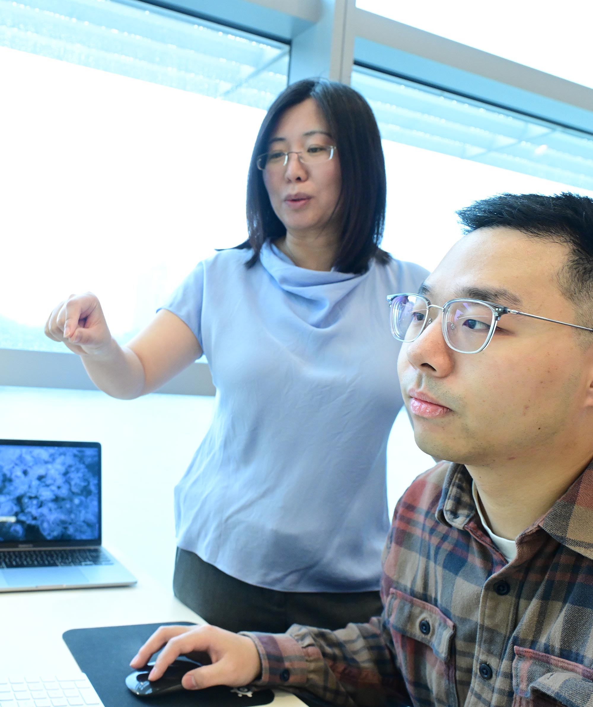
“I’m very interested in how these small proteins operate within our innate immunity,” says Li Zhao, associate professor and head of Rockefeller’s Laboratory of Evolutionary Genetics and Genomics. Zhao studies how new genes can suddenly emerge from noncoding sequences of DNA and how that helps drive evolution. Recently, researchers were surprised to find that some of these mysterious young genes are subsequently conserved across species, leading them to suspect they may in fact serve a widely useful function: protection from pathogens. To find out, Zhao is homing in on tiny chains of amino acids—known as micropeptides—that lurk within this dark matter.
This much scientists know: Once a pathogen infects a person, certain very short antimicrobial peptides rush to the infection site to help clear a pathogen and related infection.
Consisting of a chain of less than 100 acids, classic antimicrobial micropeptides detect features, such as negative charges on the membrane of the pathogen, to kill them. Others located on the membranes of host cells serve as scaffolds for immune signaling. But how many immune-related peptides remain unidentified? And do these previously unidentified micropeptides contribute to innate immunity in the same way as known ones, or do they behave differently? Could some of these novel peptides function outside of well-understood pathways, potentially revealing new mechanisms or alternative biological processes? To answer these questions, Zhao has set out to create an atlas of the types and functions of these novel micropeptides, which have been dramatically undercounted and largely unannotated.
Zhao is developing theories about how some such micropeptides come to be, but, “from a very simple perspective we still don’t know how many exist, what they mean in terms of biological response, and what kind of pathways they are regulating, especially in non-model species,” she says. The reasons are threefold.
“I’m very interested in how these small proteins operate within our innate immunity.” Zhao
First, these short proteins are so small that they often escape detection in mass spectrometers that more easily spot their larger cousins, macropeptides. Second, micropeptides evolve very rapidly, so their possible functions and roles in biology are often overlooked. Lastly, they aren’t transcribed or translated when there is no infection. So, in order to detect them, scientists need to infect cells or an organism to determine which proteins play a role in defense.
To create the atlas, Zhao and her team are studying a number of species belonging to the common fruit fly, or Drosophila, whose only defense against pathogens is innate immunity. Insects also represent a huge proportion of the planet’s animal species—about 80 percent—and are exposed to a plethora of pathogens. Because a human body’s first line of defense is similar, researchers can use what they learn from flies to better understand the mechanism in people.
With a combination of lab experiments, such as RNA sequencing infected flies and computational work, including machine learning, Zhao and three members of her lab have thus far identified hundreds of new antimicrobial and immune-related micropeptides. They’re currently working on a manuscript to describe these findings
From there, says Zhao, her team will move on to looking at genes in the adaptive response, which can illuminate the role of micropeptides in human health even more. That knowledge, along with the newly identified peptides, will be valuable in devising preventative measures and treatments for ailments ranging from those caused by novel virus strains, like bird flu, to difficult-to-treat infections, such as methicillin-resistant Staphylococcus aureus, or MRSA. Their research may also provide new insights into why some individuals are more susceptible to infections. By Sara Goudarzi
Making Magic with Cryo-EM
A new method offers a clearer picture of viral infections
For more than two decades, Hironori Funabiki’s Laboratory of Chromosome and Cell Biology has studied the way chromosomes are partitioned during cell division and the genetic changes this process creates, or what he calls “the evolution of the living system.” This work has greatly contributed to our understanding of genetic disorders, birth defects, and tumor progression.
But recently, he’s expanded his repertoire in a new direction. Because when a few researchers in his lab started discussing ways to improve the cryo-electron microscopy (cryo-EM) technique they were using to create 3D models of molecules’ structures, they ended up creating a method with major implications for studying not only chromosomes but also other small, complex molecules—especially viruses.
Their novel technique, called magnetic isolation and concentration cryo-EM (or MagIC-cryo-EM), improves the technology dramatically, allowing scientists to analyze highly diluted samples. This enables structural studies for biomolecules that are difficult to produce in their natural environments, offering a real boon to scientists investigating infectious diseases.
Cryo-EM has been a game-changer for those studying biological structures in recent decades, but the issue of sample loss has limited its use, says Yasuhiro Arimura, a former research associate at Funabiki’s lab and first author on an eLife study about the new technology. In the typical cryo-EM process, a filter is used to blot away excess liquid before imaging takes place, and much of the sample sticks to that filter, leaving only small amounts for the electron camera to capture.
Previously, this loss meant researchers had to either find molecules that were already abundant or reassemble an abundance of molecules in an artificial setting. With viruses, this compromise is particularly tricky because if researchers are studying a viral protein in a test tube, it may act differently than it would when attacking a human cell, making it difficult to predict the virus’s real-world behavior.
The goal, says Arimura, was to find a new method that would allow researchers to analyze tiny samples so they can focus in on their target protein interacting with another cell. “What we envision is to capture a cell invaded by a virus, and then catch the moment that the viral or host protein binds to viral DNA or RNA,” he says.
“This method will be very useful for infectious disease biologists, immunologists, and other structural biologists trying to solve a structure.” Funabiki
Mapping the 3D structure of a viral protein complex precisely as it enters a cell suddenly opens a door to devising new therapies or fresh strategies for preventing infection, Arimura says. Cryo-EM has been used to study the structure of viruses including Zika, SARS, influenza, and SARS-CoV-2, the virus that causes COVID-19. But studying the infection process is often difficult, and developing a much clearer picture of the host-virus interaction is urgently needed.
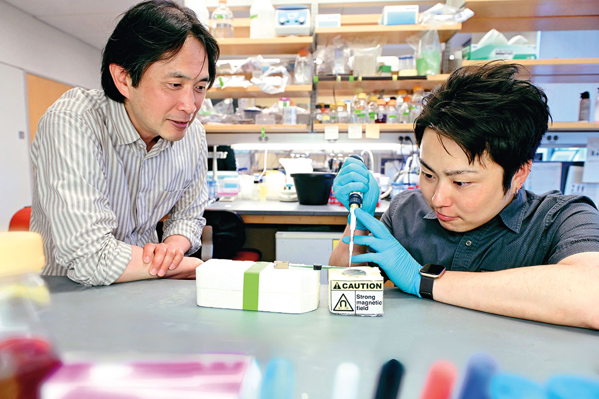
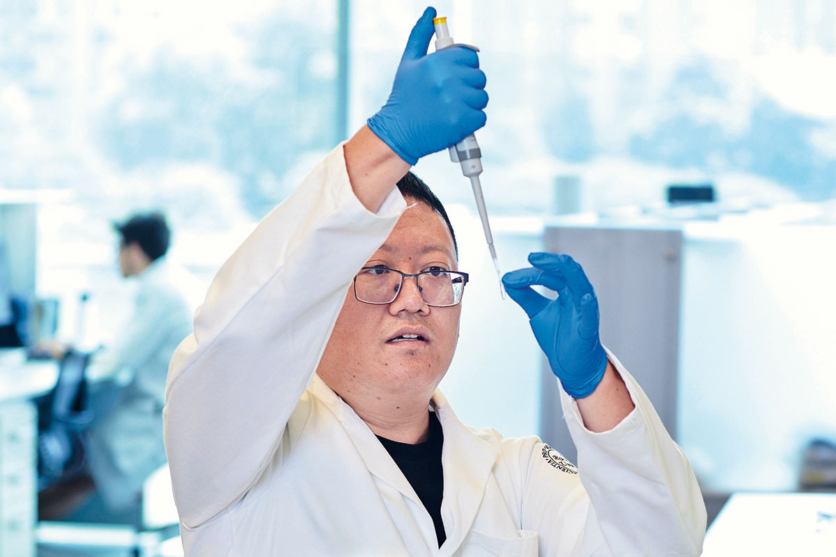
Arimura, now an assistant professor leading his own lab at the Fred Hutchinson Cancer Center in Seattle, came up with the idea for MagIC-cryo-EM while at Rockefeller. Along with his fellow postdoc Hide Konishi and Funabiki, Arimura was determined to clear roadblocks hindering their research.
Cryo-EM’s need for large sample sizes meant that researchers often used the eggs of the African clawed frog, which are big enough to facilitate the study of cell division. Interestingly, Funabiki notes, frog sperm entering an egg represents a rare example of a cell naturally acquiring DNA from outside its system, similar to a virus entering a human cell—exactly the kind of action during which traditional cryo-EM routinely failed to capture the desired molecular structures.
Arimura says he and Konishi tweaked their cryo-EM process repeatedly before their 15th version succeeded. Now, Funabiki says, researchers can use it to study a host of biological processes. For his part, Funabiki plans to leverage the group’s vast experience in chromosome inheritance in the study of how cells respond to foreign DNA.
“This method will be very useful for infectious disease biologists, immunologists, and other structural biologists trying to solve a structure,” Funabiki says. “We’ve already gotten lots of requests for collaboration.” By Abigail Abrams
Every Gene Everywhere All at Once
Tracking gene expression across millions of cells could lead to new ways to fight infections—and more effective gene therapy.
Ever tried to do 20 million things at once?
For a team of scientists studying cell behavior at Rockefeller, that’s just a day at the office. Using a technology known as EasySci, the researchers have unlocked a method to study which genes are activating at any given moment in more than 20 million cells at a time. The team previously harnessed the technology to radically revise our understanding of the aging process. Now, its inventor, Junyue Cao, who is the Fisher Center Foundation Assistant Professor and head of the Laboratory of Single-Cell Genomics and Population Dynamics, is adapting this powerful tool to the study of infectious diseases.
What a cell actually does within the body is determined by which parts of its DNA instruction manual are opened, read, and converted into the proteins that carry out its functions. If a cell is producing antibodies, interferons, interleukins, histamines, or any other proteins known for fighting infection, EasySci can now detect it. If a cell has been infected by a virus, EasySci can show the viral proteins replicating inside the cell. While the technology doesn’t offer a way to address those infections, it can tell researchers exactly where to start hunting for therapeutic targets.
This knowledge is crucial, as viruses are famously picky about which cells they infect. HIV, for example, only infects white blood cells with a specific protein, known as CD4, on their surface. COVID-19, on the other hand, is less selective but prefers cells in the respiratory system that express ACE2. Even within a population of a single virus, there are subtypes that differ slightly. These small distinctions can make a major difference in which host cells are ultimately infected. Thus, “If you can identify a virus’s targets, then you can design targeted approaches to block it from infecting those specific cell types,” Cao explains.
This ability to recognize compromised cells has already sparked a range of inventive uses. Rockefeller colleague Alexander Tarakhovsky, in collaboration with Anne Schaefer, a director at the Max Planck Institute for Biology of Ageing in Germany, is employing it to probe a vital question: Why do disease-induced cellular and organismal phenotypic states linger long after the body has cleared an infection? Tarakhovsky, a pioneer in the study of how viruses mimic our epigenomic gene regulation, now wants to understand how virus-induced modulators might be reprogramming the brain’s immune cells. Doing so could identify mechanisms driving neuronal changes supporting sickness behavior that persists beyond the original illness, as with long COVID.
“If you can identify a virus’s targets, then you can design targeted approaches to block it from infecting those specific cell types.”Cao
In this way, EasySci may yield fundamentally new insights into the damaging consequences of viral infections as well as ideas for how to treat infections themselves. But Cao is also excited about a flip side: using the technology to help viruses infect cells. While it may sound counterintuitive, gene therapies use nonpathogenic viruses as a vector to deliver genes into cells to change the way they operate. In these techniques, a modified virus is loaded with the desired gene and injected into the body. The virus then enters the target cell and tricks its host into producing the desired protein. This technique can be used to replace faulty genes, like in sickle cell anemia, or even to train immune cells to attack cancers and other diseases.
The promise of this strategy is enormous, but attempts thus far have been underwhelming due to a central difficulty that Cao’s technology is well-tailored to tackle: delivering the right gene to exactly the right place at the right time. By David Schultz