Why we need weirdos
Illustration by Nathan Eames

Forefront

Why we need weirdos

Illustration by Nathan Eames

roundworms are not known for their personalities. But as it turns out, even microscopic organisms can have an independent streak.

Rockefeller’s Cori Bargmann, the Torsten N. Wiesel Professor, has shown that genetically identical C. elegans worms, including those that have been raised in perfectly identical environments, can behave quite differently. In experiments published in Cell, her team used cameras to document every movement made by 50 worms searching for food. While most worms adhered to a standard foraging pattern, a few took the road less wiggled, departing significantly from the typical route. The scientists concluded that neural development involves a certain element of randomness—neither nature nor nurture completely determines behavior. (Read more about Bargmann’s work in “Deep secrets,” here.)

Data meter

Data

The maximum speed of a C. elegans worm is approximately 0.4 millimeters per second.

They also found a way to influence worm eccentricity by tinkering with the animals’ neurochemistry—specifically, by shutting off their serotonin production. Groups of C. elegans that lacked this chemical also lacked renegades: Every individual foraged the same way, in perfect synchrony.

Besides being boring, uniformity can be dangerous to a population. “From an evolutionary point of view, we can’t have everyone going off the cliff all at once, like lemmings,” says Bargmann. “Someone’s got to be doing something different for a species to survive.”

Sterile conditions
Photo by Matthew Septimus

On Campus

Sterile conditions

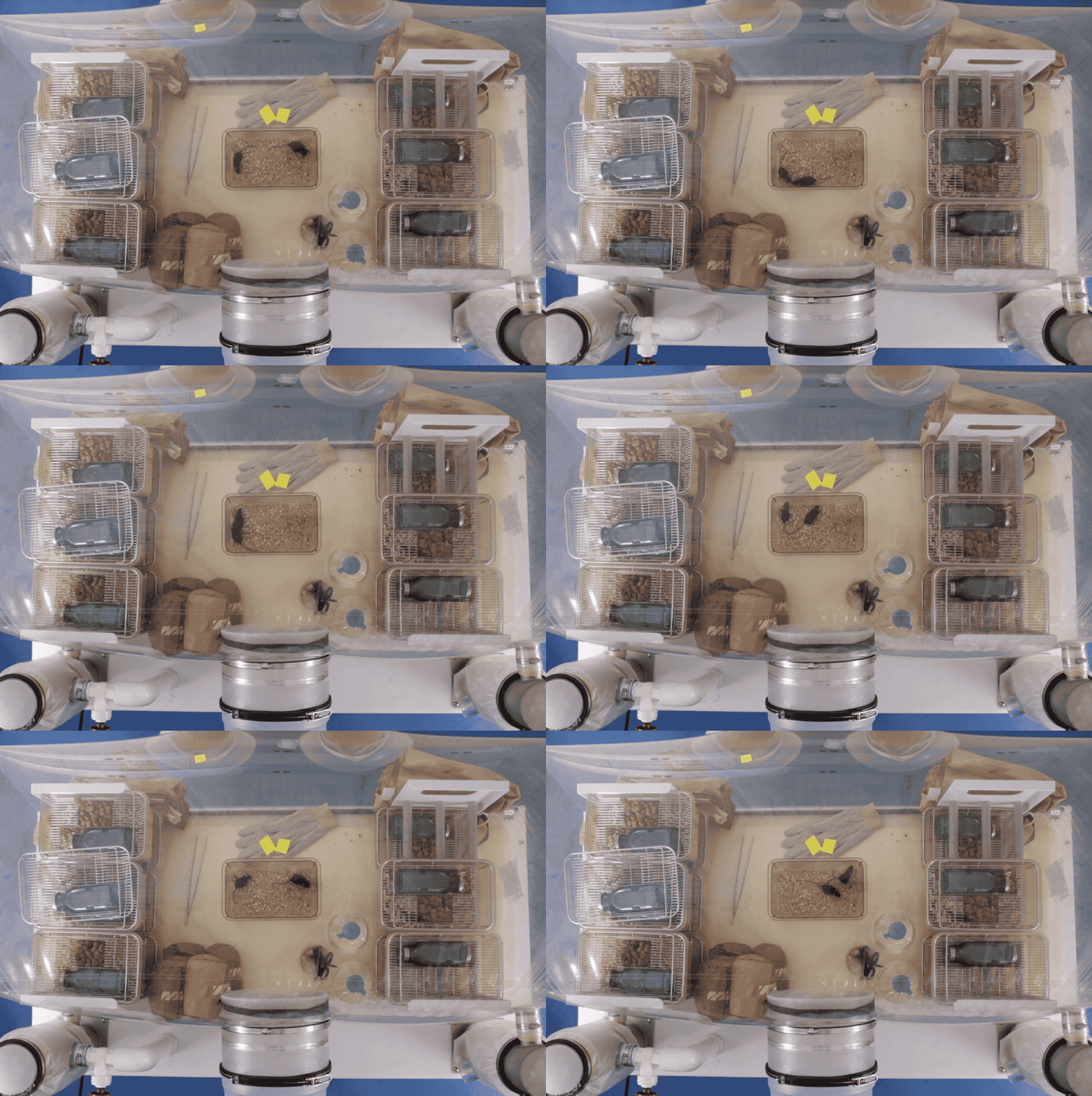
Photo by Matthew Septimus

They breathe filtered air, drink sterilized water, and eat autoclaved foodand they’ll go their entire lives without encountering a single bacterium or virus. Kept in special plastic bubblesaseptic isolatorsthese germ-free mice are the subjects of experiments, led by Daniel Mucida, to better understand the interactions between bacteria and the immune system within the gut. If we can understand how immunity and tolerance work in the absence of pathogens, Mucida says, we’ll know more about how they work in their presence.

Bad news for cancer cells and their cronies
Tavazoie is developing treatments to prevent the spread of cancer. Photo by John Abbott.

Forefront

Bad news for cancer cells and their cronies

Tavazoie is developing treatments to prevent the spread of cancer. Photo by John Abbott.

cancer cells are notoriously stubborn. When not replicating uncontrollably, they evolve new tactics to pursue their tumorous tumult. In keeping with this reputation, these malign actors have not yet surrendered in the face of immunotherapy, a new class of treatment that aims to combat cancer using the body’s own immune system.

Although researchers are optimistic about the future of immunotherapy, the treatment has yet to realize its potential—it currently works in only a slim minority of patients. One reason it often fails, it seems, is that cancer has found cellular allies within the immune system itself: tiny traitors known as myeloid-derived suppressor cells (MDSCs).

Cajoled by tumors, MDSCs stop other immune cells from doing their jobs, thereby protecting cancer cells and rendering immunotherapy ineffective. “We predicted that if we could find a way to kill MDSCs, it would lead to the activation of beneficial immune responses,” says Rockefeller’s Sohail Tavazoie, the Leon Hess Professor.

This calculation holds up, according to results from a recent study published in Cell. When Tavazoie’s team used a drug to eliminate the problem cells in mice, the intervention reduced the animals’ MDSC levels and boosted their immune powers. And the researchers obtained similarly promising results when they proceeded to test the drug, called RGX-104, in a small group of human subjects: Like the mice, human patients on RGX-104 experienced heightened immune activity as their MDSC counts fell.

Tavazoie and his colleagues will be launching a larger study to evaluate the drug’s effectiveness against various forms of cancer.

Bloodwork, working harder
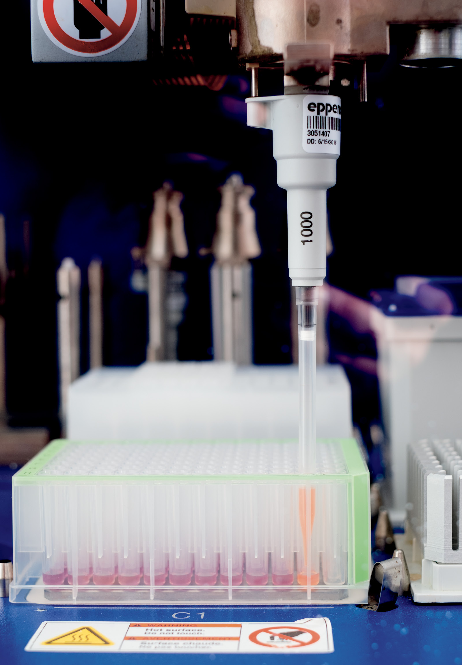
A robotic system used to process blood samples. Photo by Mario Morgado

Forefront

Bloodwork, working harder

A robotic system used to process blood samples. Photo by Mario Morgado

few people particularly enjoy having blood vacuumed out of their veins. Still, we regularly submit to clinical blood tests because, we presume, the extracted sample will alert doctors to looming disease, risk factors, or other health changes.

There’s a lot that these tests can’t tell us, however. Many medical conditions don’t leave a chemical trace, or biomarker, in the blood—at least not one that conventional techniques can decipher. Researchers in the lab of Thomas Tuschl have therefore devised a new method that widens the net of information captured in a vial of blood.

Blood Test

Data

Number of blood tests ordered by doctors in the U.S. every year.

The technique, detailed in the Proceedings of the National Academy of Sciences, involves isolating extracellular RNA, or exRNA, which cells throughout the body shed into the blood. These molecular scraps may betray medically significant details about the tissues they came from—for example, exRNA originating from the heart might be analyzed to determine the presence or progression of cardiac disease.

Tuschl and postdoctoral associate Klaas Max plan to further develop the strategy, which they hope will vastly expand the number biomarkers available for various medical uses. “This technique has enormous potential for detecting disease processes and discovering new abnormalities,” Tuschl says.

This sexy protein may help reduce mosquito populations
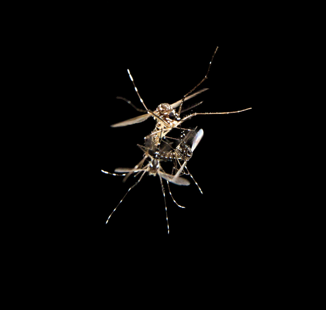
Aedes aegypti mosquitoes mating in flight. The species transmits human diseases including Zika and yellow fever. Photo by Alex Wild

Forefront

This sexy protein may help reduce mosquito populations

Aedes aegypti mosquitoes mating in flight. The species transmits human diseases including Zika and yellow fever. Photo by Alex Wild

mosquito sexual partners do not exchange sweet whispers or expensive jewelry. But male Aedes aegypti mosquitoes do leave their mates with a parting gift—a protein known as HP-I. It may not be the most romantic of presents, but HP-I can leave a lasting impression: Transferred along with semen, the protein stays in the female’s system for two hours following copulation.

Research associate Laura Duvall was curious about how HP-I affects the behavior of female mosquitoes. She and her colleagues in the lab of Leslie B. Vosshall, the Robin Chemers Neustein Professor, discovered that females receiving the protein from a mate will later spurn the advances of a second beau. If, however, a female copulates with a mutant male that doesn’t make HP-I, she will happily entertain new suitors. The protein’s function, the researchers concluded, is to discourage female promiscuity.

More than a peek into the sex lives of insects, this research, published in Current Biology, may ultimately yield public-health benefits, especially in regions affected by bug-borne illnesses such as malaria, dengue, and yellow fever. Since HP-I seems to curb females’ sexual appetite, some version of this protein could potentially be used to limit the reproduction of mosquitoes and the diseases they spread.

How we started speaking
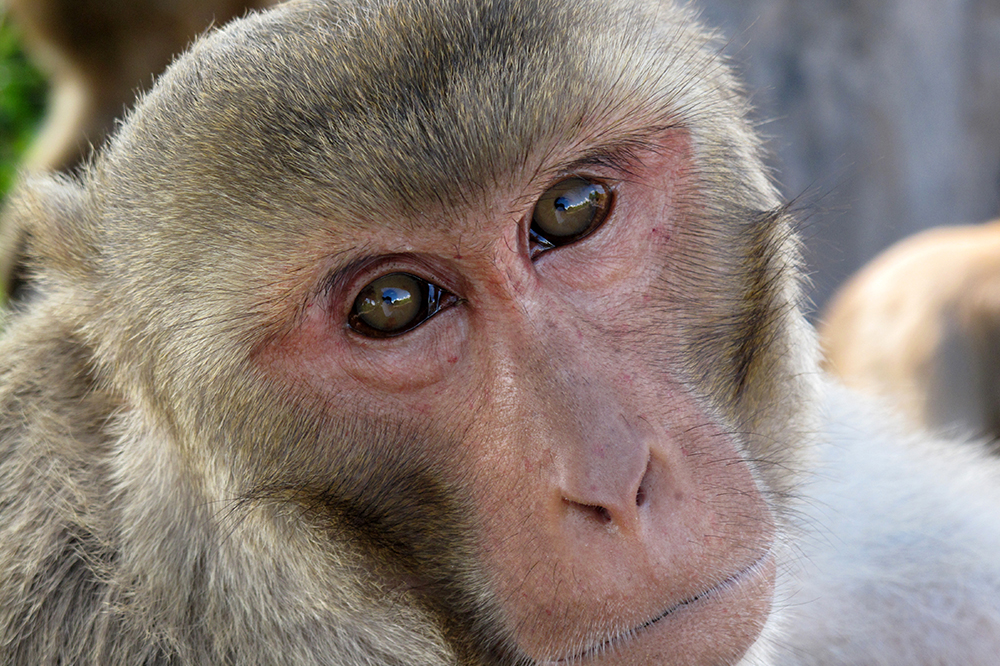
Photo by Stephen Shepherd

Forefront

How we started speaking

Photo by Stephen Shepherd

monkeys don’t talk, but they excel at body language. Facial movements, such as the friendly lip smack, are especially expressive—and they may provide clues about the origins of human speech.

Freiwald Speech

Data

Human speech may have arisen anytime between 50,000 and 2 million years ago. It’s hard to tell precisely since words don’t fossilize.

In a recent experiment, described in Neuron, Winrich Freiwald and his colleagues observed rhesus macaque monkeys as they watched videos of other monkeys, simulating face-to-face interaction. Brain scans showed that when the monkeys smacked their lips to engage with an on-screen peer, a particular brain region lit up. This part of the macaque brain resembles Broca’s area, which is known to be involved in human speech—suggesting that verbal communication may have evolved from monkey mouth movements.

Lost and found: neurons with potential healing powers
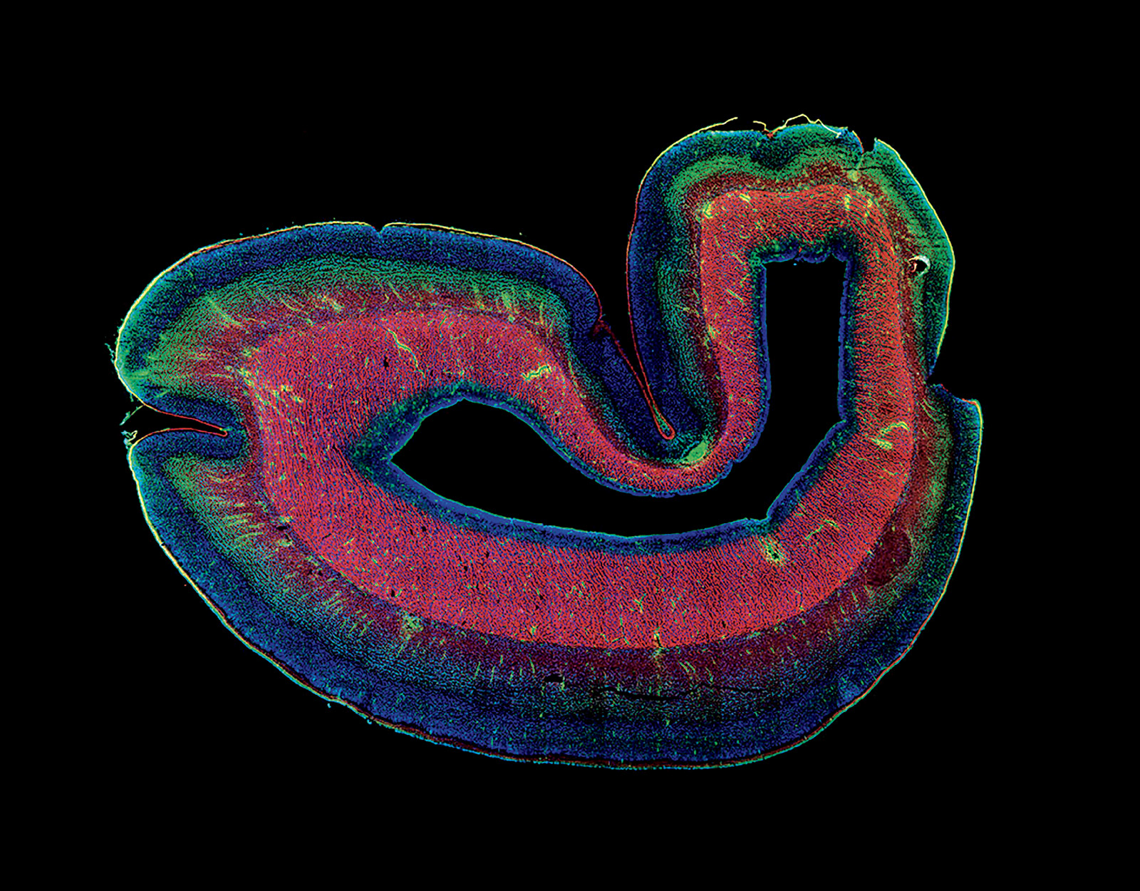
The embryonic subplate (green) sits directly below the brain’s outermost layer, the cortex, during development. Until it disappears.

Forefront

Lost and found: neurons with potential healing powers

The embryonic subplate (green) sits directly below the brain’s outermost layer, the cortex, during development. Until it disappears.

the disappearance of an entire brain region should be cause for concern. Yet, for decades, scientists have calmly maintained that one brain area, the embryonic subplate, simply vanishes during the course of human development. Recently, however, a team of Rockefeller scientists had reason to question that assumption.

“The understanding was that the cells of the subplate just die out,” says Ali H. Brivanlou, the Robert and Harriet Heilbrunn Professor. “But we hypothesized: What if these cells are not dying? What if they’re just moving to a different level of the brain’s cortex—becoming part of the cortex?”

Embryo

Data

A 12-week-old human embryo grows 15 million new neurons per hour.

Indeed, when Brivanlou’s team used a stem cell–based approach to test this idea, they saw little evidence of subplate cells biting the dust. Instead, these cells tend to mosey away from their original location, nudged along by the expression of a protein involved in neural migration. The experiments, described in Cell Stem Cell, showed that the subplate eventually moves to brain’s cortex, where its cells enjoy long and productive careers as deep projection neurons, vital to a number of cognitive processes.

The researchers also found that, with some tinkering, they could prompt subplate-like stem cells to mature into projection neuron subtypes of their choosing—a technique that could potentially become a medical strategy to replace specialized neurons lost to neurodegenerative disease.

“Alzheimer’s, Lou Gehrig’s, and Huntington’s disease all kill off specific types of deep projection neurons,” says postdoctoral associate Zeeshan Ozair. “And our research has shown us how to generate these neurons directly.”

How to fight a virus? Ask the body’s youngest cells
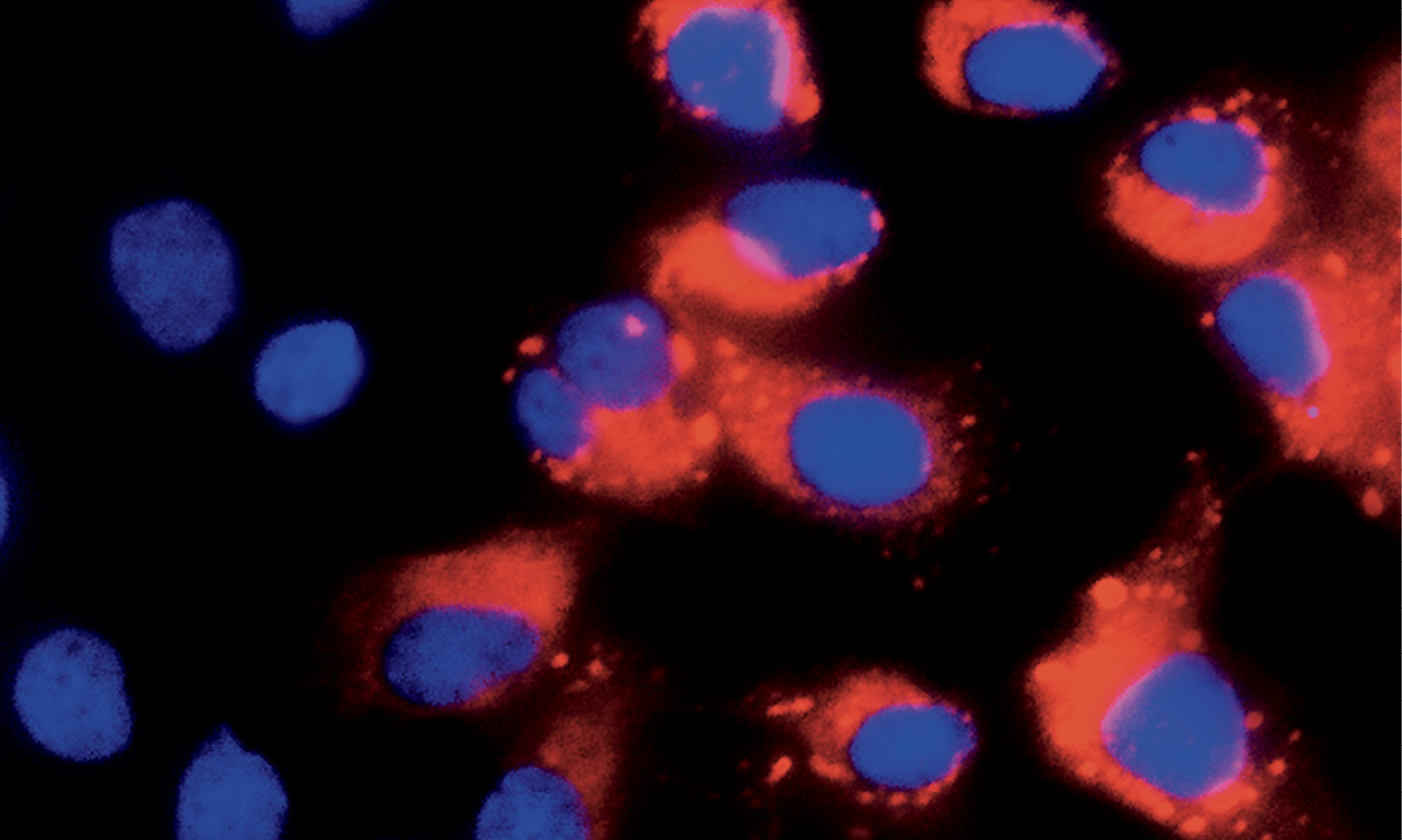
Stem cells lacking protective genes are vulnerable to attack by viruses such as dengue (red).

Forefront

How to fight a virus? Ask the body’s youngest cells

Stem cells lacking protective genes are vulnerable to attack by viruses such as dengue (red).

stem cells are the most precious of the body’s assets. Newly born and full of potential, they can grow up to be any other kind of cell—skin cell, heart cell, neuron. And, being rare and vital, stem cells receive thorough biological coddling. For example, the body has special mechanisms to protect them from the most dangerous viruses, making stem cells naturally immune to pathogens such as HIV and dengue.

“That just makes sense,” says Rockefeller’s Charles M. Rice, the Maurice R. and Corinne P. Greenberg Professor in Virology. “Because stem cells are pretty important, the body would want to be especially protective of them.”

Recently, Rice’s lab elucidated what this cellular caretaking entails: Stem cells constantly express antiviral genes, which help kick-start an immune response. By contrast, adult cells must employ these genes more prudently, switching them on only when a virus is around. These findings, described in Cell, help explain how juvenile cells stay safe, and may also lead to new insights into the defense mechanisms of older cells.

Addiction: It’s all in  your head
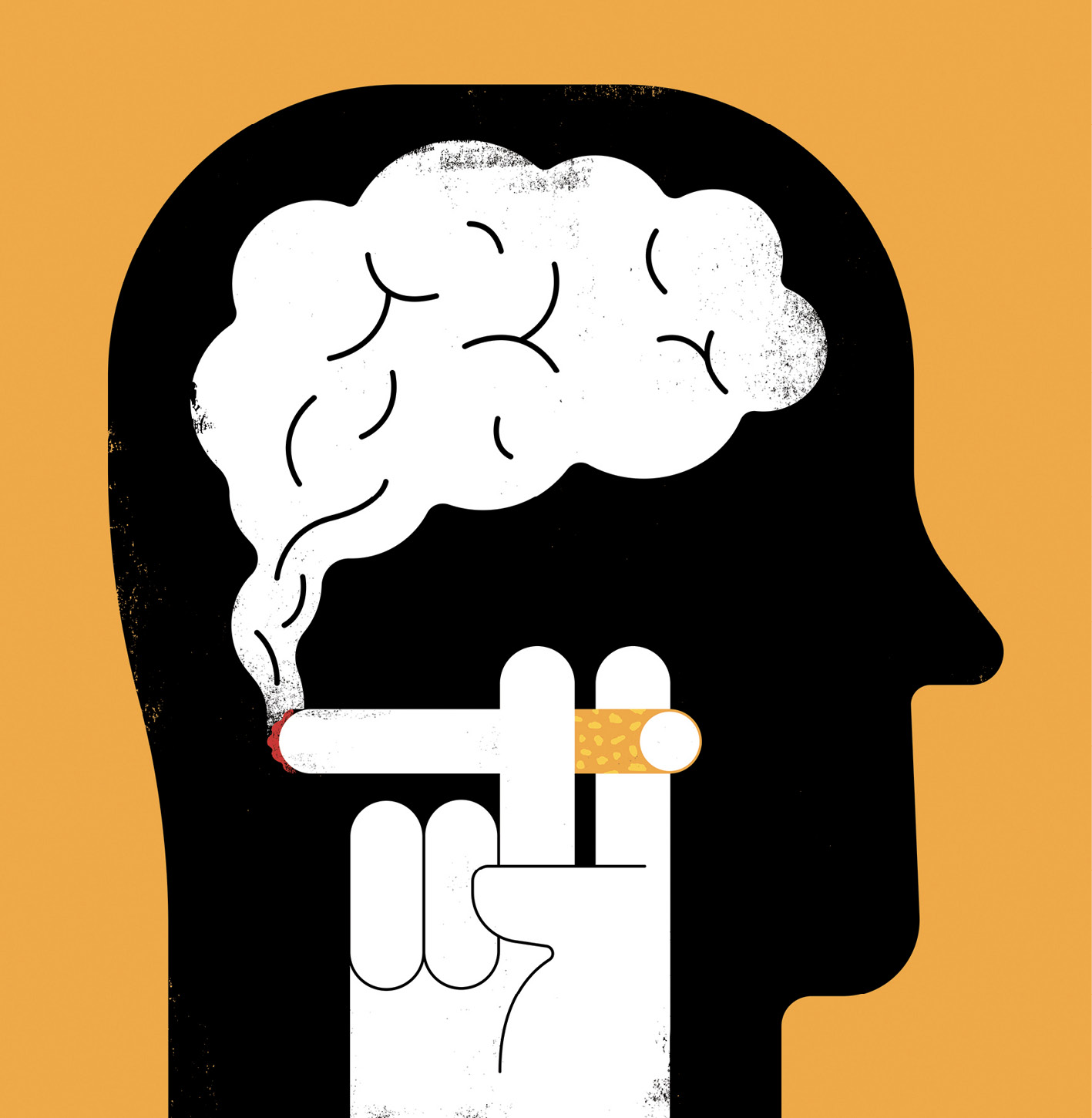
Illustration by The Project Twins

Forefront

Addiction: It’s all in your head

Illustration by The Project Twins

tobacco is the third hardest substance to quit, after cocaine and heroin. Yet smoking would be far less addictive if it weren’t for Amigo1 neurons, a newly defined class of brain cells.

In fact, the brain has a built-in aversion to nicotine: When it detects the chemical, it sends a “yuck” message to a little-studied brain region known as the interpeduncular nucleus. In a recent study, published in the Proceedings of the National Academy of Sciences, Rockefeller researchers found that, in nicotine-addicted mice, Amigo1 neurons start to produce chemicals that dilute this message.

“If you are exposed to nicotine over a long period, you produce more of the signal-disrupting chemicals, and this desensitizes you,” says Ines Ibañez-Tallon, a scientist in the laboratory of Nathaniel Heintz, the James and Marilyn Simons Professor. “That’s why smokers keep smoking.”

A Lasker in the making
Photograph by Mario Morgado

Snapshot

A Lasker in the making

Photograph by Mario Morgado

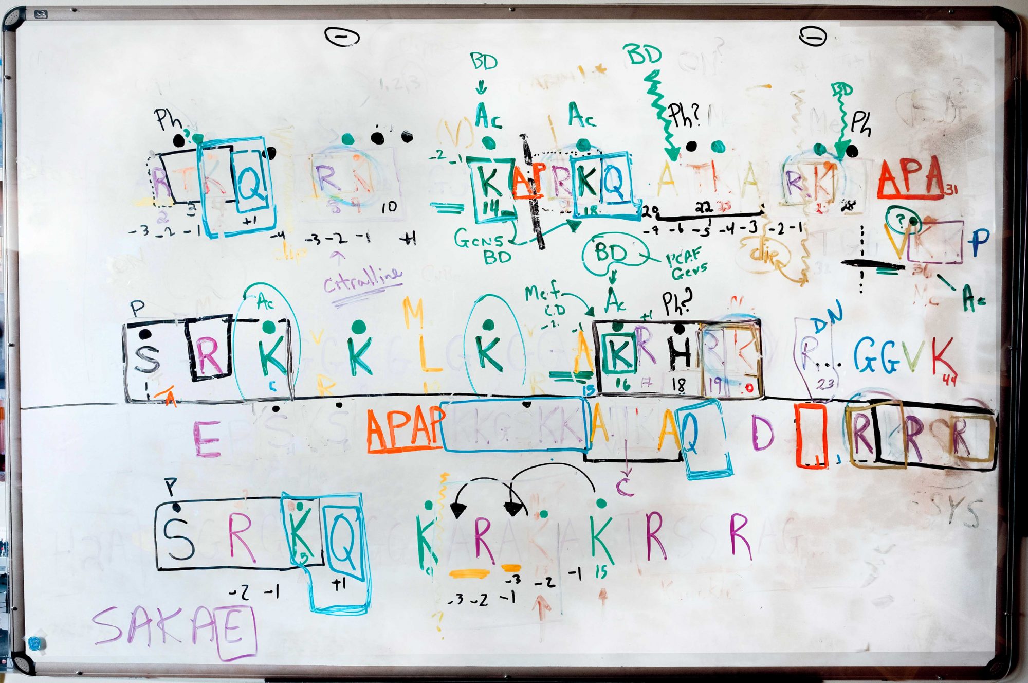
when c. david allis moved to Rockefeller from the University of Virginia in 2003, he brought a few ideas with him. In his first Rockefeller “chalk talk,” Allis laid out the details of his histone code hypothesis, which suggested an entirely new way of thinking about genes that eventually would inform almost every field in biology.

Histones—proteins that glom together to form spools around which DNA is wound—control access to specific sections of the genome. The focus of Allis’s work is on histone “tails,” which hang off the spools like loose pieces of thread. Over the years, his lab mapped dozens of proteins that make up those tails, and methodically recorded how they respond to specific enzymes to turn genes on or off. Through his research, Allis, who is the Joy and Jack Fishman Professor, has been able to confirm and refine his initial theory of gene expression; moreover, he has drawn connections between the proteins that manage histone modifications and specific diseases including heart disease, autism, and cancer. This fall, Allis received an Albert Lasker Basic Medical Research Award, one of the most prestigious honors in science, for his work.

The 15-year-old whiteboard notes from that original talk, now preserved behind Plexiglas to prevent smudging, still hang in his office.

A molecular behemoth, meticulously mapped
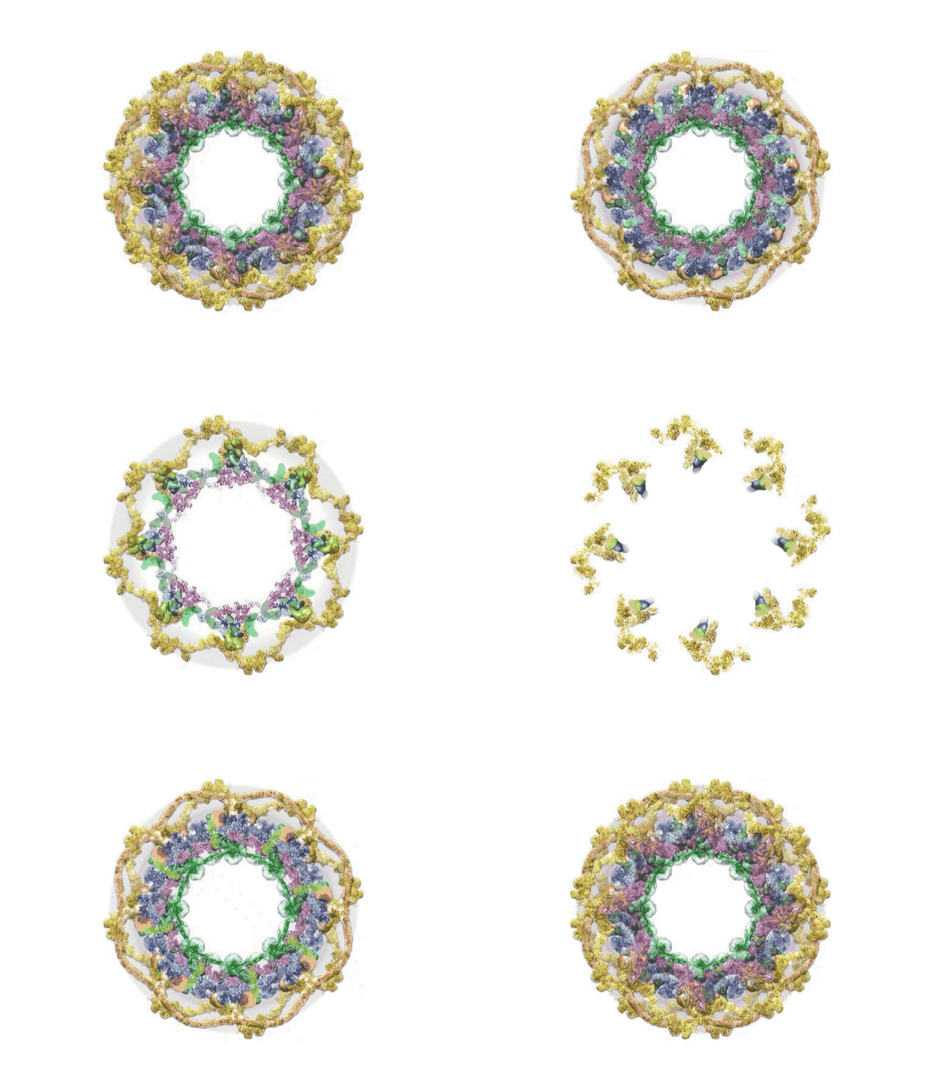
A long-anticipated map showing how the 552 pieces of the nuclear pore complex fit together.

Forefront

A molecular behemoth, meticulously mapped

A long-anticipated map showing how the 552 pieces of the nuclear pore complex fit together.

it’s hard to think of a more grandiose molecular fabrication than the nuclear pore complex, an ornate portal connecting a cell’s inner and outer compartments. The pore complex occupied a special place in the heart and mind of the late Günter Blobel, who spent decades of his life scrutinizing it (read more in “I still feel the vibration,” here). Presumably, Blobel would have been thrilled to hear the news reported in Nature in March, just a month after his passing, of Rockefeller scientists issuing the first complete blueprint of the massive structure.

It took scientists in the labs of Michael P. Rout and Brian T. Chait more than 20 years, and a medley of methods, to study the 552 components of the pore complex in yeast, and figure out how they all fit together. “In the end, we used everything we could lay our hands on, brought the results together, and integrated them into a single structure,” says Chait, who is the Camille and Henry Dreyfus Professor.

Optical tweezers
Photograph by Matthew Septimus

Gadget

Optical tweezers

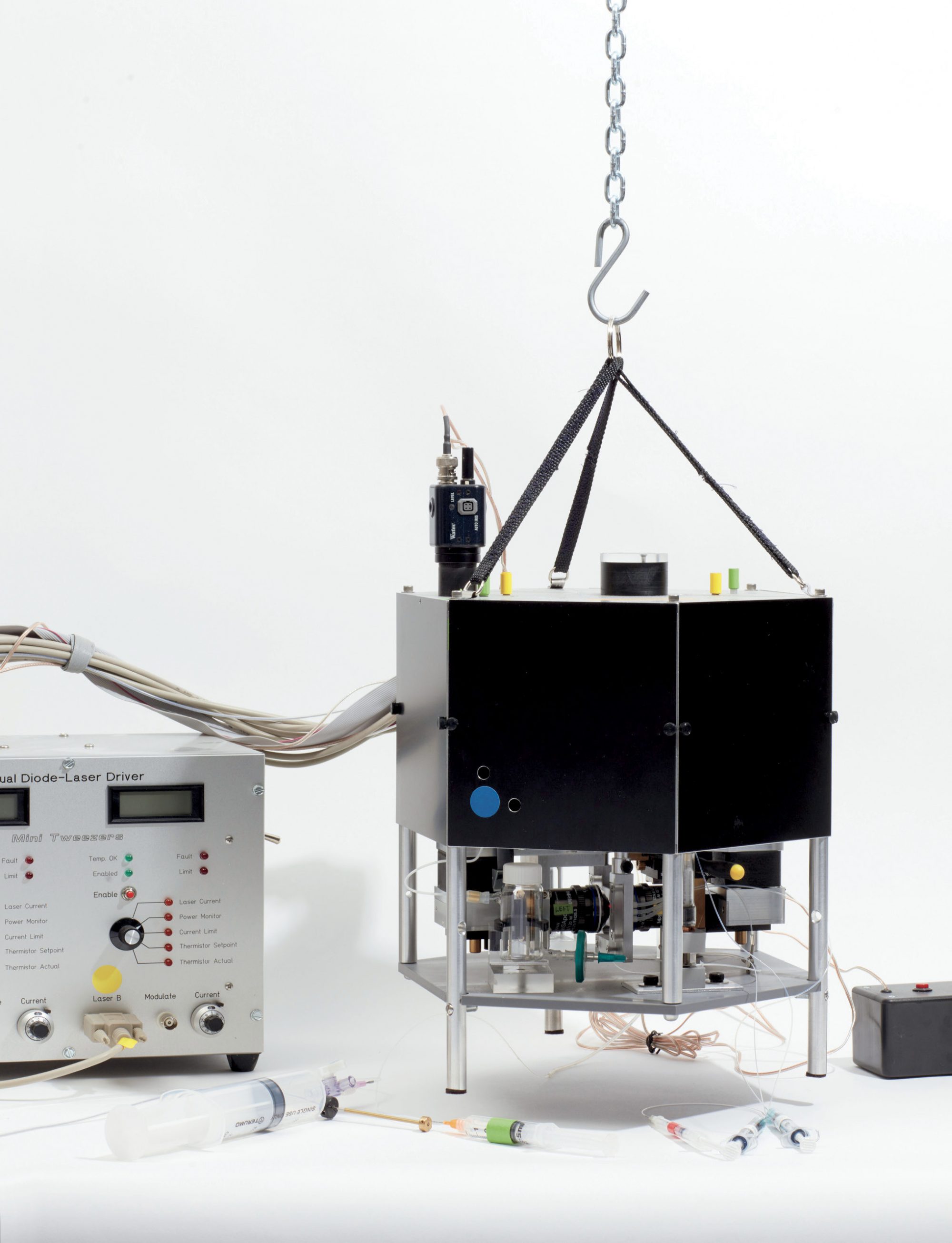
unraveling dna is harder than it sounds, at least if you want to be precise about it. You need to pull, gently but consistently, until the two strands peel apart. You need something that can hold the DNA firmly—forceps are around a million times too big—and a way to move it in precise sub-nanometer increments.

In Shixin Liu’s lab, they use optical tweezers. Specifically, a machine built by a retired UC Berkeley scientist in his garage based on tools developed some 40 years ago. The device relies on a clever combination of engineering, biochemistry, and physics.

First, DNA strands are bound to tiny, specially coated glass beads that are attracted to highly focused light. One bead is held in place with suction, and the other is “trapped” by a laser beam. By maneuvering the laser, the helix can be stretched until, eventually, it unravels.

In living cells, terrible things can happen when DNA loses its shape. Cells may mutate, die, or turn cancerous. By measuring the mechanical properties required to manipulate DNA, Liu’s group hopes to learn more about the physical rules governing genome integrity and gene expression.