Feature
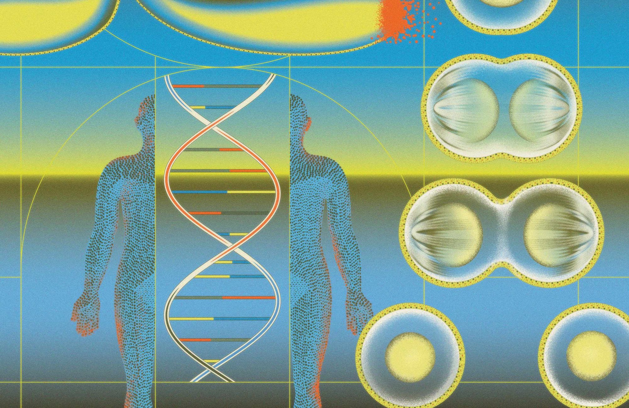
The Machinery of Life
Scientists are studying the molecular machines that read, copy, repair, and package DNA. Their findings could change how we prevent and treat diseases, including cancer.
Right now, in nearly every cell of your body,microscopic machines are copying your DNA. Clusters of proteins glide down each strand of genetic material, racing to assemble a full replica of your genome before the cell divides.Six billion base pairs are duplicated in about eight hours.
At the same time, other molecular machines are repairing DNA that’s been damaged by sources including ultraviolet light and pollution, reading the genetic code to produce proteins, and reorganizing how DNA is packaged to control which genes stay active and which fall silent. In every cell, DNA is under constant management by dozens of tiny machines that copy it, fix it, read it, and protect it.
These machines keep us alive. When they falter, the consequences are severe: Genetic mutations that go unrepaired or genes that are inadvertently turned on or off can accelerate aging and trigger cancer, spiral into neurodegeneration, or give rise to devastating inherited diseases.
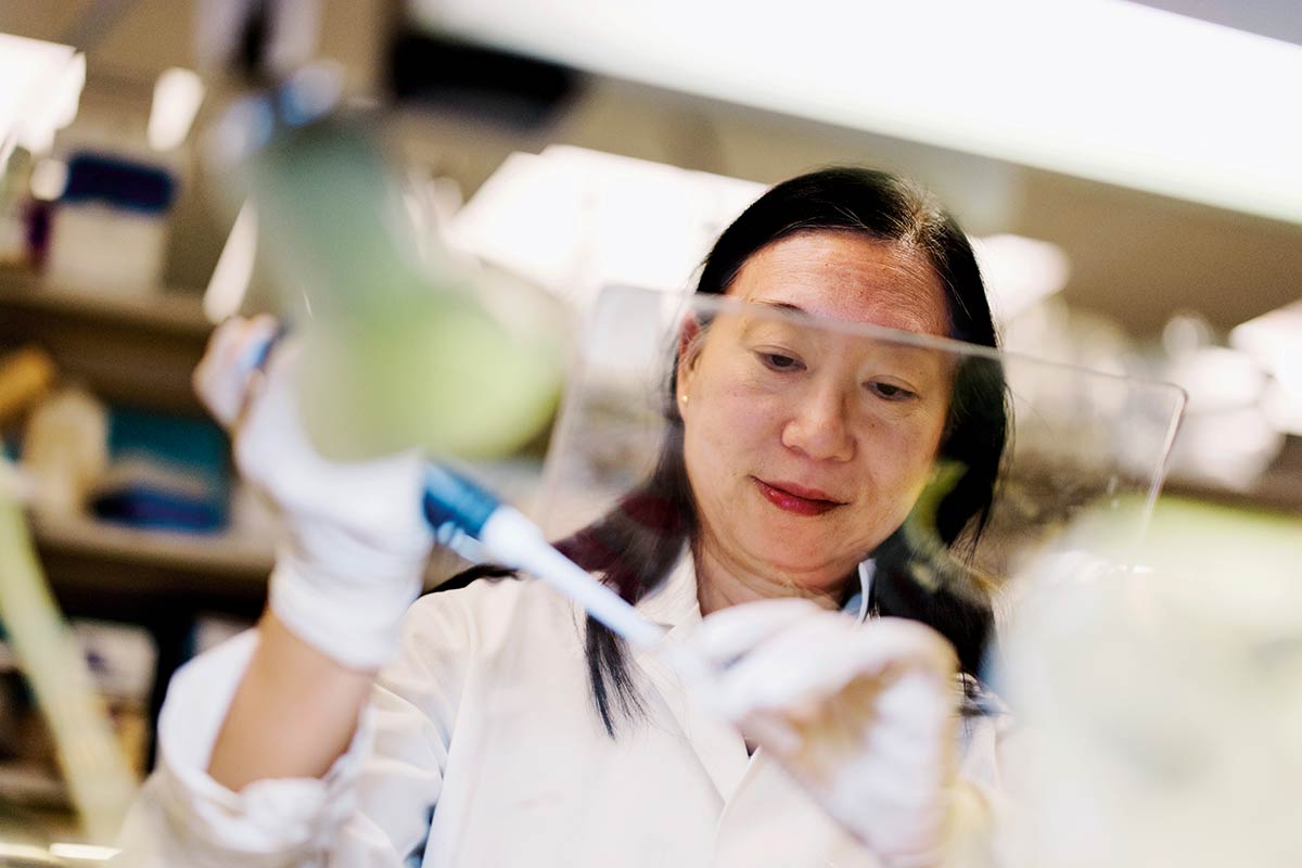
“DNA repair and maintenance are absolutely crucial for life,” says Agata Smogorzewska, the Skoler Horbach Family Professor and head of Rockefeller’s Laboratory of Genome Maintenance. “Even partial deficiencies in these pathways can disrupt cell function and lead to disease, including cancer.”
For more than eight decades, Rockefeller has been mining the mysteries of these minuscule machines. In 1944, physician-scientist Oswald Avery and his Rockefeller colleagues Colin MacLeod and Maclyn McCarty were the first to demonstrate that DNA carries genetic information, a discovery that launched the entire field of molecular genetics. Now, researchers across the university are dissecting how cells process, protect, and use that genetic material. What they’re learning could one day lead to targeted therapies that may allow us to repair damage in cancer cells, aging tissues, and genetic disorders.
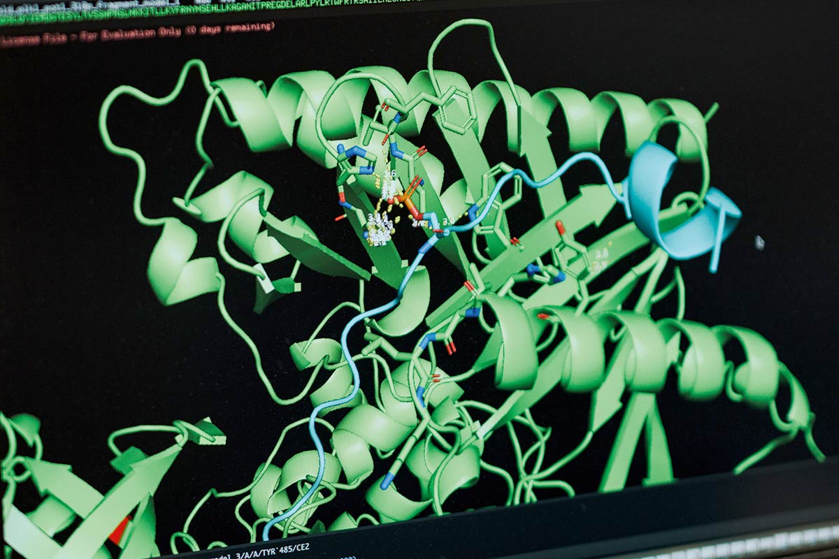
⬤ Genetic Copy Machines
The replication fork is the region where the DNA double helix separates into two single strands so that each can be copied; it’s one of the most dynamic sites in the cell. At each fork, proteins move along the exposed templates, reading the genetic sequence and synthesizing new DNA at an average rate of about 50 nucleotides per second in human cells. This is where DNA replication happens—it’s also a major source of potential errors that must be corrected by a wide variety of repair proteins, or corrected “on-the-spot” by the replication machinery itself. If a replication or genomic repair protein becomes mutated, the outcomes can be lethal.
If scientists could fix damaged genes or remove cells with harmful mutations, they could help cells stay better protected when DNA replication and repair systems fail or become overwhelmed. In fact, DNA replication and repair lie at the heart of many diseased states. For example, when abnormal amino acid is present within the replicative DNA polymerases, that can render them far less accurate than normal, leading to mutations during cell division that are associated with cancer.
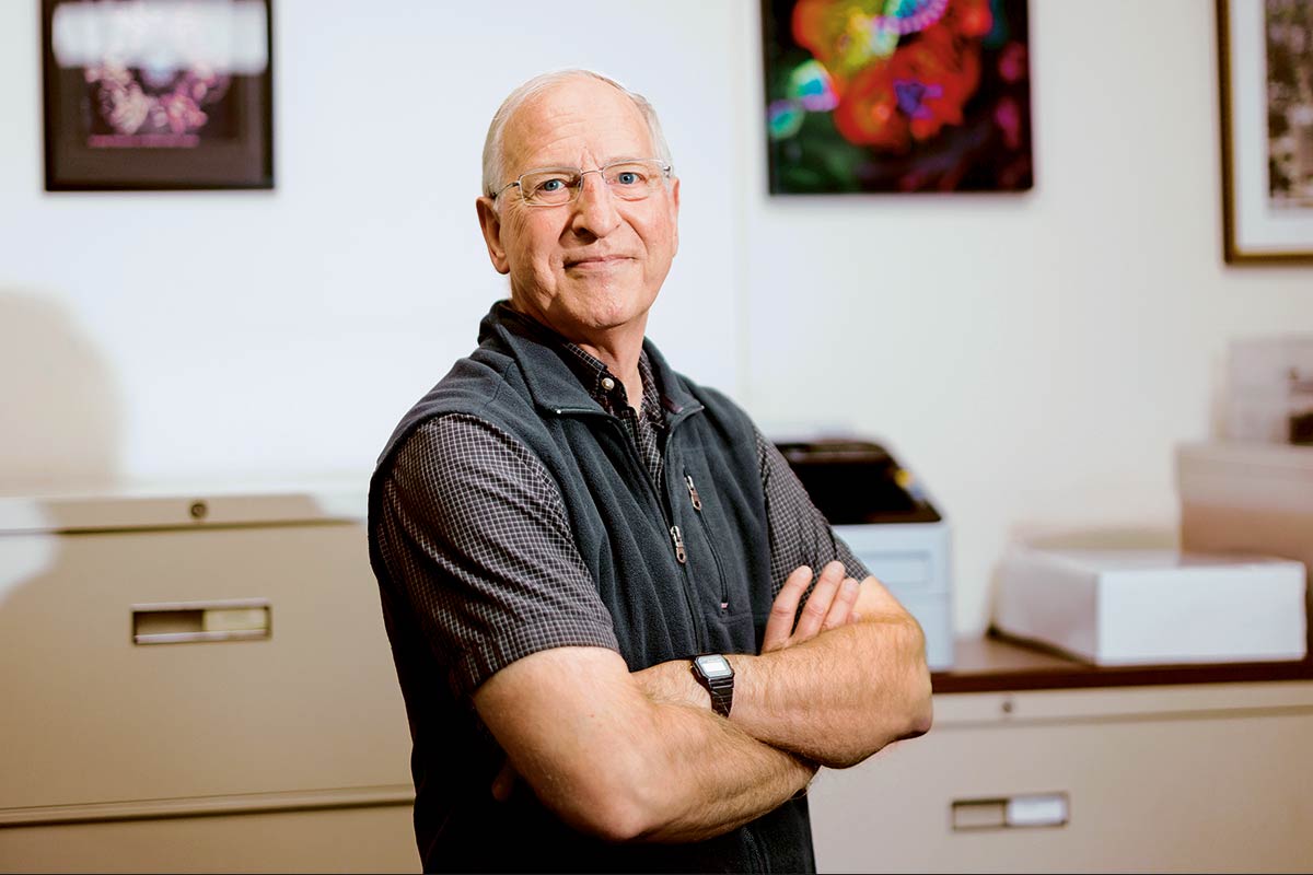
Polymerases aren’t the only potential vulnerabilities; there are numerous DNA repair factors and other DNA replication proteins that, when mutated, can also contribute to cancer and a wide range of other diseases, including conditions associated with premature aging.“There’s much we have yet to learn, but understanding all the complexity of how DNA replication and DNA repair machinery works will ultimately lead to new medical breakthroughs,” says Michael O’Donnell, head of the Laboratory of DNA Replication and an investigator at the Howard Hughes Medical Institute.
O’Donnell has spent more than 30 years patiently teasing out the staggeringly complex dance of DNA—what mechanisms govern its replication and what factors keep errors in check—to explain not just what missteps lead to rapid aging and cancer, but also neurological disorders. Early on in his research, he discovered the first protein known to encircle DNA; later studies have shown that it plays a role in all cellular life. This ring-shaped protein, known as proliferating cell nuclear antigen (PCNA), slides along DNA, physically tethering other proteins to the DNA to help them stay attached until their task in replication or repair is complete. O’Donnell and other scientists have also revealed how the PCNA ring gets loaded onto DNA—another protein complex called replication factor C (RFC) opens the ring, positions it around the double helix, and snaps it shut. Then, according to every previous model, RFC drifts away.
But O’Donnell suspected that if he took a deeper look at RFC, he’d find more to this story. Recently, he teamed up with Shixin Liu, head of the Laboratory of Nanoscale Biophysics and Biochemistry, to study the details of how and when RFC and PCNA interact with DNA. They stretched a single strand of DNA between two microscopic beads held in place by lasers. Then they added RFC that glowed green and PCNA that glowed red. Under the microscope, the researchers spotted a dot of yellow—red and green merged in the same place—moving along the strand of DNA. The new data suggested that RFC remains bound to PCNA throughout the entire replication process, traveling down the DNA together as its own complex.
 “There’s much we have yet to learn, but understanding all the complexity of how [this DNA] machinery works will ultimately lead to new medical breakthroughs.” ⬤ O’Donnell
“It’s not a result that a lot of people were expecting,” says Gabriella Chua, a postdoc in O’Donnell’s lab and former graduate student in Liu’s lab. “RFC was thought to do its job and leave the scene—and now we know that it remains bound and plays a functional role.”
Work like this has helped PCNA emerge as a potential drug target for cancer. Because it serves as a hub that binds dozens of different DNA replication and repair proteins, it acts like a master switch. Block PCNA’s interactions, and you could simultaneously prevent cancer cells from copying their DNA and repairing chemotherapy damage.
⬤ The DNA Fix-It Crew
Children born with Fanconi anemia face devastating complications from birth: missing thumbs, malformed bones, and heart defects requiring early surgery. Later in childhood, their bone marrow begins to fail, and they develop aggressive mouth and throat cancers usually only seen in lifelong smokers. Their health challenges are all because of a single molecular failure: Their cells cannot repair a type of DNA damage known as interstrand cross-links, where the double helix’s two strands become glued together when they should separate for copying.

The damage that causes this disease may sound severe, but it’s actually routine—it occurs constantly, in every person’s DNA. Ultraviolet light from the sun, air pollution, grilled meat, and alcohol chemically alter DNA. Even normal everyday activities in our cells, such as turning food into energy or controlling gene activity, generate reactive molecules that attack the genome multiple times each day.
“The only difference between the DNA damage that is accumulating in people with Fanconi anemia and in the rest of us is that most of us have a functioning system for repairing the damage,” Smogorzewska points out.
Cells have evolved an intricate surveillance system—molecules that detect damage and dispatch repair crews to fix problems before they become permanent, or stop cells from dividing when they’ve accumulated too much genetic wear and tear. In most people, this fix-it crew keeps us healthy most of the time. But when these systems fail, it has huge implications: newly introduced genetic mutations can drive cancer, accelerate aging, trigger neurodegeneration, or derail development before birth.
Smogorzewska has identified some of the 23 genes that, when mutated, lead to the inability to repair DNA cross-links, resulting in Fanconi anemia. Thus, studying these genes, she says, not only could lead to treatments and earlier diagnoses for patients, but offers a window into how DNA repair works when it’s functioning properly.
Understanding how to make the cross-link repair pathway more active or less active could also inform therapies. Turning up the pathway would be a boon to people with Fanconi anemia—their cells could begin repairing the damage so it doesn’t accumulate. Many labs are working on gene therapy to correct the underlying genetic problem. A huge challenge there, however, is ensuring these therapies reach cells all over the body.
For those patients with normal DNA repair, on the other hand, turning down the Fanconi anemia pathway could help eliminate tumors: Cancer cells would die when unable to repair cross-links induced by chemotherapy drugs. But at the moment, selectively targeting this pathway remains extremely challenging. Many dividing cells in the body rely on cross-link repair machinery to survive daily DNA damage. Turning the pathway down in stem cells could trigger mutations leading to cancer.
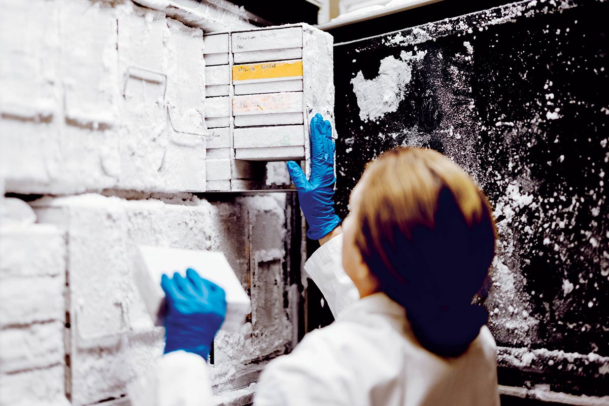
Another avenue for therapeutic intervention in Fanconi anemia patients is preventing the damage from occurring in the first place. To that end, learning more about the cells’ own sources of DNA damage could inform strategies for reducing or blocking this damage before it occurs. While the Smogorzewska lab is working on the disease-driving processes, they’re simultaneously developing ways to halt and even reverse cancer at early stages of its development in Fanconi patients. Thus far, her group has identified biomarkers that they will test in a cancer prevention trial that her lab will soon be starting.
 “The only difference between the DNA damage that is accumulating in people with Fanconi anemia and in the rest of us is that most of us have a functioning system for repairing the damage.” ⬤ Smogorzewska
DNA cross-links, of course, represent just one type of obstacle the replication machinery encounters. Other types—breaks in the DNA itself or extra chemical groups stuck to a strand—must also be repaired or avoided while DNA is being duplicated, to prevent the errors from being passed to new cells. Often, the copying machinery pauses while a repair mechanism swoops in to fix the problem.
O’Donnell and Liu have studied this machinery with their single-molecule approach. Their data suggested that when the replication machinery encounters a lesion, one of its key components—the Cdc45-Mcm2-7-GINS (CMG) helicase—doesn’t simply stay at the replication fork. Instead, it leaves the fork yet stays attached to the DNA. Once the damage is fixed, the molecule reenters the fork and reassembles the machinery to continue copying. It is a feat, they found, made possible by CMG’s ability to shapeshift on DNA.
O’Donnell’s team also uncovered how Polymerase Alpha, the enzyme that initiates each new DNA strand, can bypass certain common types of damage on its own. It’s a strategic trade-off that keeps replication moving when repair isn’t fast enough, allowing damage to be fixed later.
⬤ Chromosome Caretakers
Some changes in our DNA are not due to the onslaught of mutagens or mistakes in DNA replication, but occur slowly with each cell division. When the machinery that duplicates DNA gets to the end of a chromosome, it stops before it has copied the last few dozen base pairs. This process gradually shortens the chromosome’s protective caps, called telomeres, which are regions composed of a repeated sequence and the specific protein complex shelterin. If the number of repeats is sufficient, shelterin can bind and protect the chromosome ends. Without shelterin’s protection, cells respond to their telomeric ends as if they are DNA breaks, which stop cell division or lead to genome instability.

When cells have undergone many divisions, the telomeres become very short, and there is no longer enough telomeric DNA for shelterin to hang on to. As a result, cells stop dividing and undergo a process called replicative senescence or die. “This is actually a great thing, because it keeps cells from dividing indefinitely,” says Titia de Lange, the Leon Hess Professor and head of the Laboratory of Cell Biology and Genetics. “In fact, we now know that telomere shortening is one of the most powerful barriers to cancer development.”
 “Anything critical to telomere length regulation may well be critical to cancer prevention too. This is a major focus of our lab.” ⬤ De lange
For this cancer barrier to work, telomeres need to have the right length at birth: not too short and not too long. If telomeres are too short, cells stop dividing sooner, and this limits tissue maintenance and repair. If the telomeres are too long, early cancer cells can undergo many more divisions before they reach the barrier, frequently leading to tumor formation. “How the length of our telomeres at birth is regulated is poorly understood,” de Lange says. “Because of the importance of this issue to cancer prevention, this is a major focus of our lab.”
On the other hand, many cancers find a way to circumvent the telomere barrier by activating telomerase, the enzyme that can add telomeric DNA to chromosome ends. “Telomerase is a great target for cancer therapy,” de Lange says. “Past worries about the effect of telomerase inhibitors on stem cells may have been unfounded. It would be wonderful if somebody found a good small molecule inhibitor of telomerase to test in preclinical models.”
⬤ Speed Controls
Copying DNA is only the beginning. To turn DNA’s instruction book into the proteins that carry out much of the molecular work in living tissues, cells rely on an enzyme called RNA polymerase II (Pol II), which was first identified over 50 years ago by Robert G. Roeder, head of Rockefeller’s Laboratory of Biochemistry and Molecular Biology. When a gene is activated, Pol II latches on to the beginning of the gene and scans the code, churning out a corresponding strand of RNA through a process called transcription that can be shuttled to protein-making machinery elsewhere in the cell.
The speed at which Pol II moves along DNA, it turns out, matters a lot. After it latches onto a gene, Pol II first crawls slowly, often pausing near the start. Then regulatory proteins kick in, propelling it into high-speed transcription mode. Near the gene’s end, it decelerates again to finish cleanly. This pacing must sync seamlessly with the machinery that processes and packages RNA. Move too fast or too slow, and RNA molecules emerge malformed or misprocessed, and the risk of genome instability increases. Failure to properly control transcription speed has been linked to developmental disorders, neurodegenerative diseases, and cancer.
“I’m often asked: As long as it can make RNA and we know how it does it, do we really care about the speed of the machine or whether it pauses?” says Liu. “We care because we know that the altered kinetics of transcription are linked to various diseases.”
“What’s really striking is how this machine functions almost like a sophisticated automobile. It has the equivalent of multiple gears, or speed modes, each controlled by the binding of different regulatory proteins.” ⬤ Liu
To learn more about how cells control the speed of Pol II, Liu recently collaborated with Joel E. Cohen, head of the Laboratory of Populations, and a group of scientists in China. Their labs built a unique platform that reconstructed an entire transcription system using purified proteins—not just Pol II, but all of the other proteins that play a role in the process at the same time. This allowed scientists, for the first time, to watch these molecules move on DNA and produce RNA in real time.
The result was an incredibly precise view of a molecular engine in action, revealing exactly when and where Pol II accelerated, paused, or shifted gears, and which other proteins helped control each transition.
“Our platform allows us to objectively assess when this machine shifts gears, and how fast it goes,” explains Cohen.
The team opened up a molecular gearbox: a hierarchy of regulatory proteins working in concert to control speed. One protein acts as the master switch, chemically modifying Pol II to unlock its activity. Others serve as accelerators, stabilizers, or brakes, each binding at specific moments to push the enzyme faster or hold it back.
“What’s really striking is how this machine functions almost like a sophisticated automobile,” Liu says. “It has the equivalent of multiple gears, or speed modes, each controlled by the binding of different regulatory proteins. We figured out, for the first time, how each gear is controlled.”
The hierarchy also reveals potential drug targets. One of the master control proteins, positive transcription elongation factor B (P-TEFb), was already known to be a promising target for leukemia and solid tumors, including breast, pancreatic, and ovarian cancers; blocking it can keep the cancer cells from expressing their malignant genes. But scientists have struggled to do so without toxic side effects on healthy cells. Understanding exactly how it fits into the larger speed control system could enable more precise therapeutics.
“Our work may offer clues for designing more specific therapeutics, and a better understanding of what could go wrong in disease,” says Liu.
⬤ Genome Architects
DNA isn’t just a long flat string of genetic code. It’s carefully wrapped around proteins called histones, which are organized into chromatin—a complex packaging system that controls which parts of the genome are active and which should be silent by determining which sections can be accessed by molecular machines. This architecture makes it a challenge for replication, repair, and transcription machineries to access the genetic code.

Viviana I. Risca, head of the Laboratory of Genome Architecture and Dynamics, has developed ways to map the organization of chromatin proteins inside cells. She has shown how changes in this architecture can alter cancer cell biology—work that could contribute to the development of new cancer therapies.
Risca’s lab is investigating the mechanism by which loss of function of proteins associated with chromatin organization—including linker histones, which help compress histone-wrapped DNA into its most compact form—can alter gene expression, which other groups have shown contributes to the emergence of blood cancers.
“The importance of linker histones in compacting chromatin in blood cells agrees very well with human patient data where linker histones are some of the most highly mutated genes in lymphoma cases,” Risca says.
These mutations can trigger a cascade of events: chromatin regulation fails, genes or stretches of repetitive “junk” DNA that should stay silent become active, and DNA repair systems cannot keep up with the onslaught of damage from all this activity on DNA. The Risca lab’s work showed that the molecular mechanism by which the chromatin regulation fails involves unfolding of chromatin throughout the genome, which has context-dependent effects on genes. As a consequence, a cell’s ability to maintain a stable identity—say, a B cell’s ability to remain a B cell—is compromised. The gene expression patterns suggest that it reverts to a more primal state that divides uncontrollably and becomes cancerous.
“Linker histones are some of the most highly mutated genes in lymphoma cases.”⬤ Risca
Risca’s work has also revealed something unexpected about cancer treatment. When breast cancer cells are treated with standard chemotherapy, most tumor cells die, but some persist in a kind of resting state known as senescence. As the cells enter senescence, Risca and Justin Rendleman, a postdoc in her lab, discovered that the cells begin expressing genes not usually active in adult tissues. These aberrant genes mark the drug-resistant cancer cells like a red flag and present a potential opportunity to target them with additional therapies that will kill them without harming healthy tissue. The Risca lab is currently exploring such avenues and working to understand how the rearrangement of tumor cell chromatin during drug-induced senescence turns on such marker genes. It’s just one example of how understanding chromatin architecture could unlock new therapeutic strategies.
The broader lesson from chromatin research is that DNA management operates at multiple levels simultaneously. Cells must not only copy DNA accurately and repair damage quickly; they must also organize the genome so that the right genes are accessible at the right times, repair machinery can reach problem sites, and transcription machinery moves at appropriate speeds.
When any of these levels fails—replication, repair, transcription, or packaging—the whole system can unravel. Understanding how they work together, and how they fail individually, is essential for developing therapies that target disease at its molecular roots.
“Just as the work of Avery, MacLeod, and McCarty ignited the molecular revolution by revealing DNA as the basis of heredity, current research at the university aimed at understanding the fundamental biology of genome maintenance will drive future therapeutic breakthroughs,” says Smogorzewska. “The impact on human health could be equally revolutionary.”