The brain on quarantine
Illustration by Katherine Lam

Forefront

The brain on quarantine

Illustration by Katherine Lam

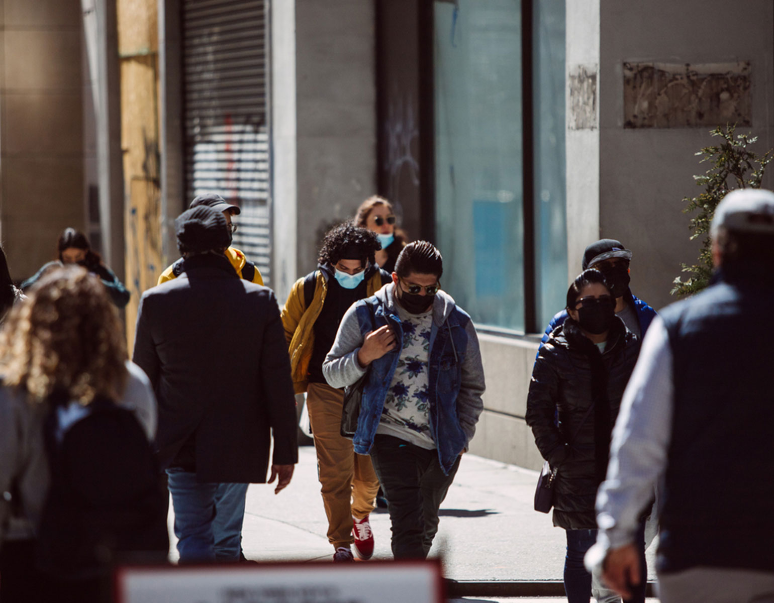
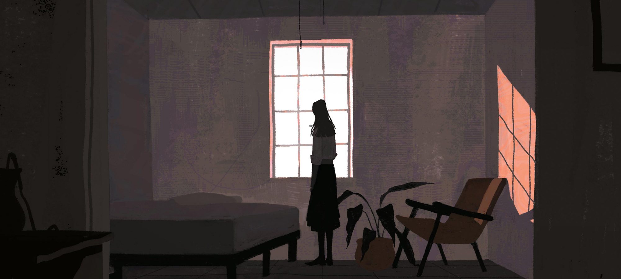
Loneliness is toxic. Not only can it make you feel sad or unfulfilled, but it has been linked to various health issues, from increased blood pressure to depression, cognitive decline, and cancer. The year 2020 was a case in point: Under shelter-in-place orders, Americans tended to be more anxious and depressed, had shoddier sleep quality, and, according to some estimates, put on more than half a pound per person every 10 days—a recipe for medical problems.

On the other hand, scientists have found that when people experience a sense of belonging—in a romantic partner, a pet, or at the local quilting club—they tend to live longer. So, what changes in the brain when we’re cut off from society?

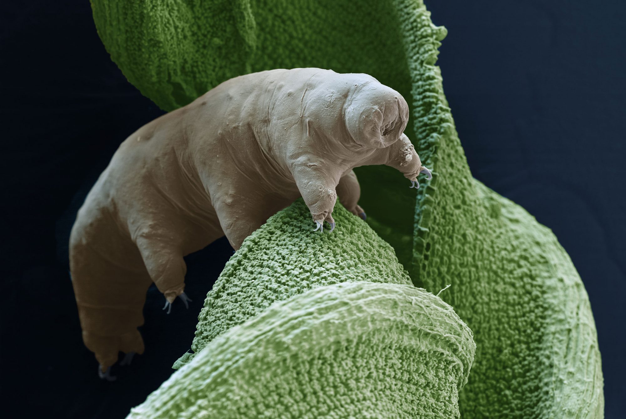
Recently, scientists went looking for answers in a somewhat unexpected model, the seemingly primitive Drosophila melanogaster. Despite its tiny brain, the fruit fly is in fact a gregarious little creature with a highly complex social life. It forages, feeds, and explores its environment in the company of peers. It engages in elaborate mating rituals that, like human wedding traditions, are passed down through generations by social learning. And according to the new research, it suffers under lockdown.

The brain on quarantine

Data

In a recent study, 36 percent of Americans reported feeling lonely “frequently” or “most of the time.” Young adults aged 18–25 and mothers with young children were among the most affected.

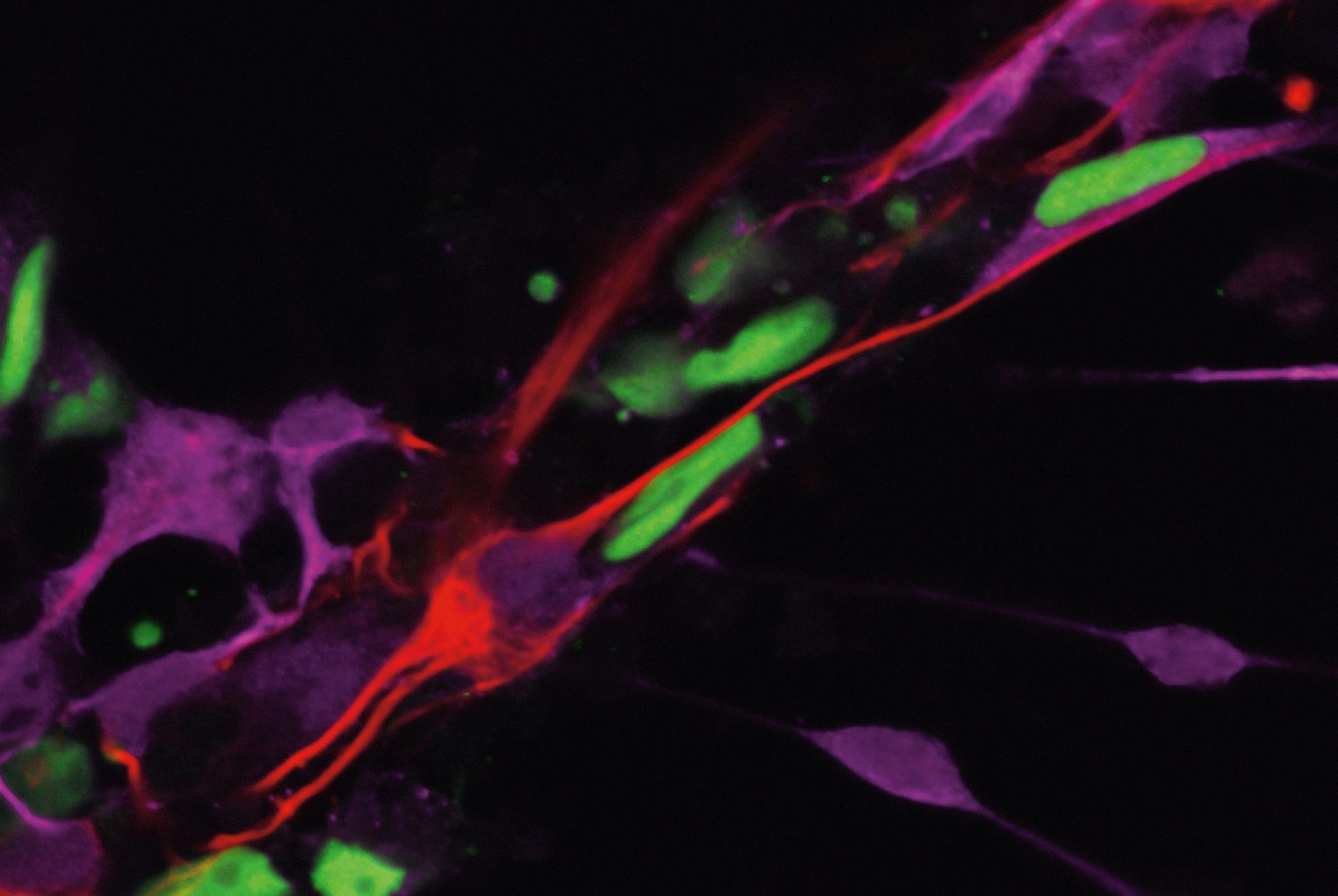
Wanhe Li, a research associate in the lab of Michael W. Young, let some flies abide on their own while congregating others in groups of various sizes. Seven days into the experiment, the solitary flies were sleeping less and eating more, just like isolated Homo sapiens. The control flies carried on sleeping and eating normally, however, as long as they had one or more companions.

“It may well be that our little flies are mimicking the behaviors of humans living under pandemic conditions,” says Young, who is Rockefeller’s Richard and Jeanne Fisher Professor and a 2017 Nobel laureate.

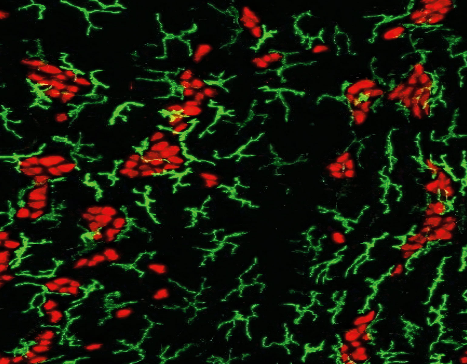
The scientists identified a small group of neurons that might be driving a fly’s loneliness response. When they shut down these neurons in genetically manipulated flies, the animals maintained normal sleep and feeding patterns, even after a week in exile. The findings, published in Nature, might inform research into the biology of social isolation in mammals, which is currently a black box.

“When we have no road map, the fruit fly becomes our road map,” Li says.

Kaleidoscopic colors
Photo by C&G Partners, Designers

On Campus

Kaleidoscopic colors

Photo by C&G Partners, Designers

For two decades, Rockefeller’s Pearl Meister Greengard Prize has been shining a light on the accomplishments of extraordinary women scientists. Now, light shines through a translucent display of the winners, installed recently in the Abby Aldrich Rockefeller Lounge. Passersby can experience a play of shifting colors as the iridescent panels catch the light at different angles­­—an effect inspired by natural phenomena like butterfly wings and oyster shells. So far, 23 panels have been etched with past winners’ portraits, and blank ones stand ready to celebrate future awardees.

How petite pedestrians evolved
The tardigrade walks on eight stubby legs, with a gait resembling that of insects 500,000 times its size. Eye of Science / Science Source

Forefront

How petite pedestrians evolved

The tardigrade walks on eight stubby legs, with a gait resembling that of insects 500,000 times its size. Eye of Science / Science Source

Tardigrades: you can freeze them or burn them. You can shoot them out of a gun at 18,000 miles per hour and even expose them to the cold vacuum of space. These dumpy micro-animals, also known as water bears, will shake it off and live to plod another day. Water bears are virtually indestructible, but that isn’t the most remarkable thing about them if you ask Jasmine Nirody, a visiting fellow in Rockefeller’s Center for Studies in Physics and Biology, whose fascination with the hardy tardigrade stems from the way it gets from point A to point B.

“No other animal of that size can walk like a tardigrade,” Nirody says.

At less than a millimeter in length, one would expect tardigrades to wiggle or thrash about, like similarly appointed roundworms or insect larvae. Instead, the tardigrade trudges through soil and water upon stubby legs, in the ponderous, bear-like gait that earned it its nickname.

“The similarities between this locomotive strategy and that of much larger arthropods like beetles and scorpions have very interesting evolutionary implications,” says Nirody, whose research appeared in PNAS last year. Among her most impactful discoveries is that water bears, like scurrying insects, can switch from a leisurely stroll to a mad dash by simply increasing the speed of a single stepping pattern. This transition is different from that of a horse raring into a gallop, for example, because it doesn’t involve swapping one movement pattern for another. And the findings could mean that a wide range of panarthropod species, from insects to water bears, share a common ancestor.

But it is also possible, Nirody notes, that the soft tardigrade lacks ancestral links to insects and other hard-shelled creatures. It may have evolved its little legs independently. That would suggest that a water bear’s tread and an insect’s scuttle are practical solutions when it comes to navigating unpredictable terrain with a small body­­—and that such solutions are repeatedly favored by natural selection.

Intestinal memories
Laboratory of Mucosal Immunology

Forefront

Intestinal memories

Laboratory of Mucosal Immunology

The gastrointestinal tract boasts the largest cache of neurons outside of the brain itself—acting like a “second brain” that, among other things, controls the body’s flow of nutrients and waste. A mighty army of T cells and macrophages protects these neurons, defending them from stomach bugs and other stressors.

100M

Data

Number of neurons in the human small intestine. The brain has at least 800 times as many.

Daniel Mucida, who heads the Laboratory of Mucosal Immunology, has shown that these immune cells learn to rally around GI neurons exposed to foodborne diseases. His lab recently demonstrated that mice suffer neuronal assault mainly the first time they are infected with salmonella or certain parasites; with a second infection, their gut neurons remain unscathed.

“We’re describing a sort of innate memory that persists after the primary infection is gone,” Mucida says about the findings, published in Cell. He suspects that some GI conditions may occur when the body fails to develop this tolerance, leaving its second brain out on a limb.

The butt of a joke, redeemed
Where are the neurons that ensure we never forget a familiar face? Illustration by Keith Negley

Forefront

The butt of a joke, redeemed

Where are the neurons that ensure we never forget a familiar face? Illustration by Keith Negley

The theory of the grandmother neuron—a single brain cell purportedly responsible for remembering specific faces, like that of your grandmother—was dismissed just a few years after it was first put forth, in the 1960s. Decades later, when Winrich Freiwald trained as a neuroscientist, people would mostly refer to it in jest.

“If you wanted to ridicule someone’s argument, you would dismiss it as just another grandmother neuron, a hypothetical that could not exist,” Freiwald says.

But the central question that had prompted the grandmother neuron hypothesis endured: What happens in the brain when we spot a familiar face, or any recognizable object for that matter? According to modern neuroscience, neurons collaborate rather than act on their own. They don’t operate like buttons on a control panel—no single neuron can produce a complex brain function all by itself. If anything, neurons are more like piano keys whose coordinated activities create endlessly intricate harmonies.

Facial recognition apparently works the same way. Some neurons process visual face data, for example, while others are tasked with storing such information. So imagine Freiwald’s surprise when his team recently discovered—well, not the quintessential grandmother neuron, but what he believes might be the closest thing to it.

In findings published in Science,the researchers used functional magnetic resonance imaging to monitor neural activity in subjects viewing a selection of photos, including portraits of individuals the subjects had previously encountered in the flesh and those they had seen only virtually, on a screen. An interesting pattern emerged within a small face-recognition area in the brain’s temporal pole region.

“The neurons responded three times as strongly to faces that the subjects had seen in real life,” says Sofia Landi, a former graduate student in Freiwald’s lab now doing postdoctoral work at the University of Washington. This could mean that our brains react differently when we see people we’ve gotten to know on Zoom, Landi says, compared to those with whom we’ve had real-life encounters.

Moreover, the experiments showed that the temporal pole neurons are unlike any other cells known to be involved in face recognition. They simultaneously behave both like sensory cells and memory cells and are hence able to connect our visual perception of a face with our remembrance of it. In that sense, the cells seem to play a role similar to that once ascribed to the legendary grandmother neuron.

The analogy goes no further, however. A temporal pole neuron doesn’t code for a specific familiar face, neither Granny’s nor that of one’s mother, boss, Nancy Pelosi, or anyone else.

“What we’ve discovered is more like a grandmother face area of the brain,” Freiwald says.

Wake up and smell the coffee
Josefina del Mármol has discovered how olfaction differs from other forms of sensory perception at the molecular level. Photo by Mario Morgado

Forefront

Wake up and smell the coffee

Josefina del Mármol has discovered how olfaction differs from other forms of sensory perception at the molecular level. Photo by Mario Morgado

Earthy, nutty, cocoa with a hint of caramel—the aroma of a perfect cup of coffee. More than 200 chemical components coalesce in carafes and demitasse cups around the world to produce that familiar scent. And the human nose happily receives the message.

But how the brain then processes this surge of olfactory information is one of the great mysteries of neuroscience. Because while millions of molecules can unite in countless permutations to form any number of unique smells, humans are endowed with only a few hundred odor receptors to sniff through it all. And unlike most sensory receptors, which bind only to specific molecules, those that detect odors must multitask among many different ones.

Theories about how odor receptors pull this off have long wafted through the neuroscientific community. Some suspected that the receptors are glad to bind to any molecule that possesses a few basic features. Others proposed that odor receptors are as selective as any receptor but are pockmarked with numerous binding sites, allowing a single receptor to interact with many different molecules at once.

Yet when Josefina del Mármol, a postdoctoral associate in Vanessa Ruta’s lab, inspected the odor receptors of an insect known as the jumping bristletail, visualizing them at atomic resolution, she found evidence for neither approach.

Instead, del Mármol and her colleagues reported in Nature that each odor receptor contains a single pocket that can form weak bonds with several different odorants. “The receptor is not selective to a specific chemical feature,” Ruta says. “Rather, it’s recognizing the more general chemical nature of the odorant.” Computational modeling revealed that one particularly hardworking receptor sports a pocket that is at once selective and accommodating—rejecting unwanted odorants while weakly binding to many others.

Ruta suspects that the findings can be extrapolated to humans. “They point to key principles in odorant recognition, not only in insects’ receptors but also in receptors within our own noses that must detect and discriminate the rich chemical world,” says Ruta, the Gabrielle H. Reem and Herbert J. Kayden Associate Professor.

Coveted, COVID-proof genotypes
Scientists suspect that some people carry mutations that protect them from ever getting COVID. Getty Images

Forefront

Coveted, COVID-proof genotypes

Scientists suspect that some people carry mutations that protect them from ever getting COVID. Getty Images

As COVID continues to find new victims, one of its biggest mysteries remains unresolved: why certain people appear to keep dodging it.

We all know of individuals who quickly lost their lives to the disease. Others had infections that triggered long COVID, a concoction of debilitating symptoms that may linger for years. Still others, including fully vaccinated people with limited exposure to the virus, caught the disease several times.

And then there are the curious cases of people with ample exposure who never got sick. Among those who shared a bed with an infected partner or those who spent months with COVID patients in the ICU, some never tested positive. Are these individuals impervious to the disease, or did they just escape it by chance?

“We want to know if there are gene variants that protect people from SARS-CoV-2 infection,” says Jean-Laurent Casanova, Rockefeller’s Levy Family Professor, who is leading a major effort to answer that question. “If there are, and we could find them, that would be huge.”

480

Data

Number of inherited mutations shown to make the immune system vulnerable to specific pathogens. So far only four mutations have been shown to protect from infections, but many more may be out there.

If mutations that prevent infection indeed exist, the researchers want to learn precisely how they stop the virus from replicating. Knowledge of those mechanisms could make it possible to develop antiviral drugs that make people less prone to catching COVID, and less likely to spread it to others.

András N. Spaan, a postdoctoral fellow in Casanova’s lab, adds that human genetics studies have traditionally focused on the type of mutations you don’t want—those that are linked to poor outcomes. “But we can learn a lot about the pathophysiology of an infectious disease by studying beneficial mutations that make the immune system better equipped to deal with it,” he says.

With an international consortium of scientists, Spaan and Casanova are recruiting participants for a clinical study aimed at discovering the genetic factors of COVID resistance. They have already heard from hundreds of people from around the world who’ve demonstrably been exposed to the virus without being infected.

To learn more about the trial, visit the COVID Human Genetic Effort at www.covidhge.com.

Our dicey future
Bacteria found in soil produce an antibiotic effective against multidrug-resistant pathogens. Photo by Jacob Arthur Pritchard

Forefront

Our dicey future

Bacteria found in soil produce an antibiotic effective against multidrug-resistant pathogens. Photo by Jacob Arthur Pritchard

Let’s face it: Public health is in a tight spot. Not only is the world still plagued by the viral pandemic; experts have long warned that, unless novel antibiotics are developed, multidrug-resistant bacteria will soon render current ones inefficient. Already, at least 700,000 people die each year from infections with strains like XDR Acinetobacter baumannii and Neisseria gonorrhoeae that don’t respond to existing antibiotics. Even colistin, long used as a crucial last option when other drugs fail, is becoming obsolete.

But hardworking scientists might be able to forestall the impending apocalypse. In January, a team led by Sean F. Brady, Rockefeller’s Evnin Professor, reported in Nature their discovery of macolacin, a natural compound that might make it possible to vanquish pathogens that don’t respond to colistin or other antibiotics. A chemical cousin of colistin, macolacin is produced by soil bacteria that live in conflict with other microbes.

When the researchers synthesized and tested macolacin, they were impressed by its potency. In cell assays, the agent killed several types of colistin-resistant bacteria, including intrinsically resistant N. gonorrhoeae; and in mice, it completely cleared away infections with colistin-resistant A. baumannii.

Both strains are common causes of infections in health-care settings, and both are classified as public-health threats of the highest level by the Centers for Disease Control and Prevention. A novel drug to defeat them would be a promising milestone on the road away from a superbug dystopia.

When bacteria self-vaccinate
Viruses (blue) latch on to a bacterial cell. The bacterium may use a combination of primordial defense strategies to destroy the invaders. Science Photo Library

Forefront

When bacteria self-vaccinate

Viruses (blue) latch on to a bacterial cell. The bacterium may use a combination of primordial defense strategies to destroy the invaders. Science Photo Library

Like most organisms, bacteria are prey to viruses—and their go-to approach for destroying the invaders is to simply chop them up. When it sees a virus, a bacterium may employ a host of immune strategies to slice up its genome using a molecular cutter called CRISPR-Cas, also the name of a popular laboratory tool.

But before engaging CRISPR-Cas, the bacterium will usually launch a first line of defense: its so-called restriction enzyme, a molecule capable of cleaving short DNA sequences. If the restriction enzyme fails to cut the virus and stop it in its tracks, CRISPR-Cas, a more sophisticated weapon, comes online. The CRISPR cutter slits the viral intruder with immaculate precision by neatly aligning it to a molecular guide targeting a specific genetic sequence. Whereas the restriction enzyme approach chops up viral DNA with the crudeness of a lawn mower, CRISPR-Cas is more akin to razor-sharp gardening shears.

Both types of bacterial defense are commonly exploited by biologists whose day-to-day chores involve manipulating DNA for various purposes—like sequencing genes, making molecules fluoresce, or creating animals with modified genomes. Restriction enzymes came into vogue in the 1970s, making it possible to cut up pieces of DNA swimming in a test tube; and a decade ago, technology based on CRISPR-Cas revolutionized bioscience by giving scientists the means to edit with high precision genomes within living cells and organisms.

But Pascal Maguin, a graduate fellow in the lab of Luciano Marraffini, remains committed to exploring the bacterial basics—and, in the process, he recently clarified how one facet of bacterial immunity operates. Working with Staphylococcus aureus, Maguin and his colleagues were able to explain why the virus-chopping strategies of this bacterium work better together than on their own. When staphylococci are protected only by restriction enzymes, their defenses are short-lived; and after a while, the research shows, the bacteria growing in the dish will start to dwindle. Maguin discovered how the two systems work in concert—segments previously clipped by restriction enzymes help the CRISPR-Cas machinery gain a foothold in the viral DNA, which it then uses to generate the molecular guide needed to put an end to the infection.

“It’s a bit like vaccination,” Marraffini says. “The restriction enzyme cuts little pieces of the virus that CRISPR will then use to mount an adaptive response.”

The findings, reported in Molecular Cell earlier this year, might not only help us understand how staphylococci defend themselves from viruses but also could make us better equipped to defend ourselves from staph—a species notorious for its ability to become resistant to antibiotics and a common cause of outbreaks in hospital settings. Last year, Marraffini’s lab published other findings showing that the bacterium uses its CRISPR-Cas system not only to fend off viruses but also to develop multidrug resistance. Understanding the system better could one day allow scientists to manipulate it with drugs to fight staph infections that respond to no other treatments, says Marraffini, who is Rockefeller’s Kayden Family Professor.

Rise of the replicons
Replicons provide a safe system to study pathogens that normally require strict biosafety measures. Photo by Frank Veronsky

Forefront

Rise of the replicons

Replicons provide a safe system to study pathogens that normally require strict biosafety measures. Photo by Frank Veronsky

It’s biosafety 101: Whatever you do, don’t let the dangerous pathogen escape the lab. It’s why much of the research on SARS-CoV-2 has been done in sophisticated and massively expensive negative-pressure laboratories, complete with air locks and HEPA filtration.

For some experiments, however, there’s a more creative approach: replicons, lab-made self-replicating viral genomes that are not infectious but otherwise identical to the real pathogen. Replicons have proved instrumental in the development of drugs for other viruses. For instance, hepatitis C replicons developed by Nobel laureate Charles M. Rice, the Maurice R. and Corinne P. Greenberg Professor in Virology, led to the creation of powerful new drugs to effectively cure that disease.

“If the virus were a race car, we made a version without wheels.”

Now, given the urgency for more effective COVID drugs, Rice and his team have created SARS-CoV-2 replicons that can be used to investigate how the virus hijacks the cell’s own machinery and how it generates new copies of itself. And, as the researchers point out in a paper in Science, replicons might make it easier to develop new drugs.

The new replicons mimic nearly every aspect of the coronavirus life cycle. Their genetic content has all the information the virus needs to mass-produce copies of itself and pack them into new virus particles, but it lacks instructions for making spikes, the proteins that enable the particles to enter and infect human cells. Once introduced to cells in a dish, a replicon makes progeny that are unable to contaminate neighboring cells.

30,000

Data

Approximate size of the SARS-CoV-2 genome measured in nucleotides, the basic components of RNA. The genomes of hepatitis C, HIV, and rhinovirus are more than three times smaller.

“If the virus were a race car, we made a version without wheels. It has the engine and all the parts that would allow the car to move, but it can’t actually go anywhere,” says Joseph Luna, a postdoc in the Rice lab who worked on the project along with research associate Inna Ricardo-Lax.

Replicons are typically created by cloning DNA sequences that can be used to make replicon RNA artificially. But the researchers realized that standard cloning methods wouldn’t work for the coronavirus, whose RNA is exceptionally long. So instead, they used a platform developed by collaborators at the University of Bern and the Institute of Virology and Immunology, in Germany, which involved assembling coronavirus genomes from smaller fragments in yeast instead of synthesizing whole genomes directly in the test tube.

Luna says scientists will be able to use the replicons to test drugs against SARS-CoV-2 and evaluate its response to neutralizing antibodies. It’s a way to speed up the science without sacrificing safety.

Metabolic mishaps
Illustration by Hoi Chan

Forefront

Metabolic mishaps

Illustration by Hoi Chan

Mitochondria keep our cellular lights on. Floating in a cell’s gelatinous interior, these bean-shaped bubbles act like its nuclear power reactors, churning out energy that drives everything we do, from replicating DNA to running marathons. Moreover, failures in mitochondrial maneuvers can be Fukushima-like, leading to the accumulation of chemically reactive free radicals inside mitochondria that may trigger cancer, neurodegeneration, or other problems.

Recently, Rockefeller scientists took a leap forward in studying such havoc, known as oxidative stress. A team led by Kivanç Birsoy, the Chapman Perelman Assistant Professor, discovered how glutathione, an antioxidant produced outside of mitochondria, enters these powerhouses to neutralize free radicals. Their experiments show that glutathione transport relies on a protein in the mitochondrial membrane whose function was hitherto unknown.

Published recently in Nature, the findings might inspire further research on aging and the various diseases linked to oxidative stress. “These conditions could potentially be treated or prevented by stimulating antioxidant transport into mitochondria,” Birsoy says.

Viral venom
Jan Erhart, Biology Centre CAS

Forefront

Viral venom

Jan Erhart, Biology Centre CAS

Of the millions of spineless creatures crawling about planet Earth, ticks may be the least loved. These minuscule, hard-bodied beasts spread many dangerous infections, including tick-borne encephalitis, endemic in Russia, China, Mongolia, and many European countries. A cousin of the viruses causing dengue, yellow fever, and Zika, tick-borne encephalitis is just as nasty as it sounds—a rampant neurological disease that inflames the brain and thwarts cognition.

10,000

Data

Number of cases of tick-borne encephalitis reported worldwide each year.

In analyzing blood samples from 800 infected people, Marianna Agudelo, a graduate student in the lab of Michel C. Nussenzweig, found that some samples contained unusual antibodies capable of neutralizing the virus. As reported in the Journal of Experimental Medicine, Agudelo and her colleagues cloned these antibodies and successfully used them to curb the sickness in infected mice. They are now working to translate their findings to humans with the goal of developing new treatment and prevention methods.

For example, a vaccine that coaxes the immune system to produce the rare antibodies on its own “would be more elegant and more focused than existing vaccines,” says Nussenzweig, the Zanvil A. Cohn and Ralph M. Steinman Professor.

Journey to the cerebellar cortex
Laboratory of Developmental Neurobiology

Snapshot

Journey to the cerebellar cortex

Laboratory of Developmental Neurobiology

About 80 percent of the brain’s neurons are packed into what is sometimes called our “little brain,” the cerebellum. During development, some reach their proper places by climbing along delicate fibers extended by glia, another type of brain cell.

To recapitulate the process, Hourinaz Behesti, a research associate in the lab of Mary E. Hatten, the Frederick P. Rose Professor, grew cerebellar neurons from human stem cells. These engineered neurons (green) knew precisely what to do when Behesti inserted them into a young mouse brain. They dutifully found their glial tracks (red) and followed them toward their next developmental destination­—the compact cellular network that will eventually become the cerebellum.

Read more about stem-cell science in “Stem cells are growing up”.

N95 recycler
Photograph by John Abbott

Gadget

N95 recycler

There was a time when face masks were considered disposable. Scientists, who needed them while working with infectious agents, would routinely toss them after each use.

This changed in 2020. As it became clear that disposable face masks would need to be reused, Thomas Huber and Alexander Epstein, members of Thomas P. Sakmar’s Laboratory of Chemical Biology and Signal Transduction, set about designing a device to decontaminate masks.

Chemical disinfectants were ruled out, as was heat, which could damage the fragile fibers that trap virus particles. But ultraviolet light—specifically UVC radiation in the range of 200 to 280 nanometer wavelengths—seemed perfect: It kills pathogens by destroying the proteins that hold them together.

UV light is a brilliant disinfectant, long used in air ducts and water filters. For Huber and Epstein, the trick was to deliver UVC deep into the layers of fiber that make up N95 masks. With 36 watts of power, and translucent reflective surfaces covering 360 degrees, the chamber that the scientists built does the job, as validated with hundreds of UV-sensitive stickers and many rounds of fit testing. The most challenging part of the design, Epstein says, was the hook from which the mask hangs. “We tried a lot of designs but they all cast shadows,” he says. “In the end we found that a bent paper clip works best.”

Several chambers, the parts for which were 3D-printed in Rockefeller’s Precision Instrumentation Technologies resource center, are stationed around campus for the community’s benefit, and they’ve been used tens of thousands of times over the past 20 months.