Experiences, memories, and the elusive element of time
Illustration by Iker Ayestaran

Forefront

Experiences, memories, and the elusive element of time

Illustration by Iker Ayestaran

The way we act and react is often informed by our past—specifically, by good or bad experiences that help us project what may transpire in the future. A classic example is the Russian psychologist who rang a bell before feeding his dog—eventually the dog learned to salivate at the sound even in the absence of dinner.

But Pavlov’s story tends to glaze over an important factor in this behavioral equation. Today’s neuroscientists have reasons to suspect that associative learning isn’t merely the outcome of a cue being linked to a reward—the order of these events matters, too.

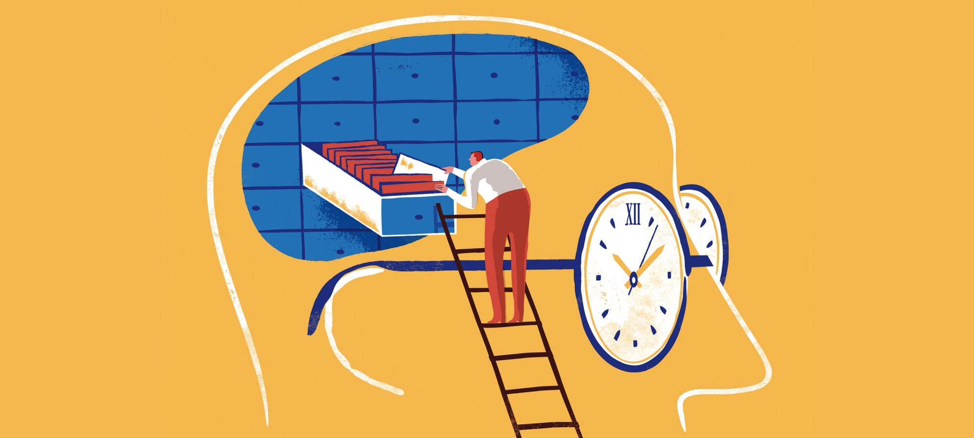
Consider a scenario where the bell normally rings after, not before, the arrival of dog biscuits. To the canine diner, the sound won’t then represent bliss, but the end of bliss; and it will presumably produce memories of sadness rather than of appetite. All of which suggests that the brain doesn’t just file experiences of different kinds in its memory bank—it somehow assigns time stamps to these experiences as well.

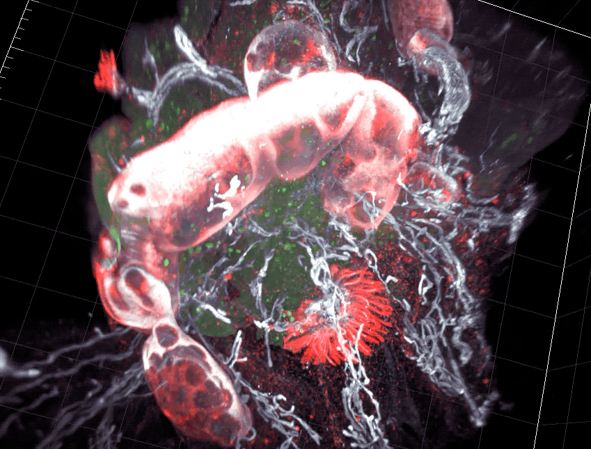
Time is a weird thing from a biological perspective, far more abstract than sounds or objects. You can’t see, hear, touch, or taste it, yet the brain seems quite capable of tracking it. Precisely how this chronicling occurs in the context of learning is something that researchers in the lab of Vanessa Ruta, the Gabrielle H. Reem and Herbert J. Kayden Associate Professor, are very keen to understand.

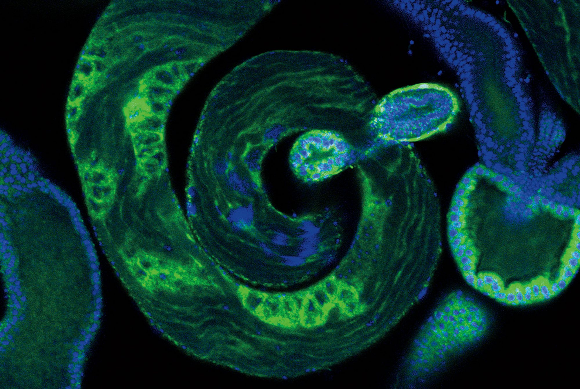
The lab doesn’t have much faith in the dog as a model system, however. For these scientists, man’s best friend is Drosophila melanogaster, the humble fruit fly. Recently, Ruta’s team set up a modern version of Pavlov’s experiment in which they exposed flies to an odor rather than to a sound; and instead of rewarding the animals with a treat, they used optogenetic technology to directly stimulate reward-signaling neurons in the flies’ brains.

The results were clear. When the flies were given a reward signal immediately after receiving a puff of the smell, they became attracted to that scent; but when the reward came before the smell, they shunned it instead. “The difference in time is only one or two seconds, yet the flies form completely opposing associations,” says graduate fellow Annie Handler.

She and her colleagues identified a set of brain cells whose activation enables the fly to know the sequence of events. In addition, they found that flies that had learned to covet the smell could quickly be retrained to detest it, and vice versa. In other words, flies are like us in that their memories are not set in stone.

“There are so many things that we could remember on a daily basis, so we hold on to the memories that turn out to be predictive; and we toss out associations that are incorrect or irrelevant,” says Ruta. “When you live in a dynamic environment—which both flies and humans do—that seems like a very good strategy.”

A historic touch

On Campus

A historic touch

Several decades of science were conducted in Rockefeller’s Smith and Flexner Halls before their interiors were gutted in a 2010 renovation. A new interactive installation, the Scientist Explorer, honors the 137 men and women with labs in the buildings—from the first who arrived in 1917 to those currently on the faculty. Occupants and visitors of today’s Collaborative Research Center, which connects the two buildings, can use the circular touchscreen to explore the work of both neighbors and predecessors.

Spermatic innovators
Photograph by Mario Morgado

Forefront

Spermatic innovators

Photograph by Mario Morgado

Sperm science has a curious past, supported at times less by fact than by the (predominantly male) imagination. In the late 17th century, when Dutch scientists caught the first glimpse of the tadpole-like cells under the microscope, they decided that each one carried within its head a miniature human being that would grow into a baby—positing that the egg cell, discovered decades earlier, played but a minor role in human reproduction.

This myth was eventually debunked, but others followed, including the still-popular idea that ejaculated spermatocytes purposefully swim toward the egg, propelled by an ancient drive to outcompete sperm from other males. In reality, these cells are much less heroic: They don’t even swim very far but passively drift across the uterus, buoyed along by soft motions in the female tissue.

In short, we have yet to see the evidence of machismo manifesting at the micro scale.

This isn’t to say sperm are completely without agency, however. They do have at least one impressive talent: an unsurpassed knack for building novel genes. In this sense, the testes are not mere sperm factories but also laboratories churning out fresh DNA content—undeniably a seminal mission given that new genes are the raw material for the evolution of species.

Recently, scientists took a major step toward understanding how nature’s attempts at innovation play out during the development of sperm. Working with fruit flies, a team in the laboratory of Li Zhao created a detailed map of DNA mutations arising in each sperm and the activity of new genes arising from those mutations.

“It offers an unprecedented perspective on a process that enables living things to adapt and evolve, and that ultimately contributes to the diversity of life on Earth,” Zhao says about the research, published in eLife earlier this fall.

Her team is interested in so-called de novo genes that emerge from scratch rather than through duplication of existing genes. The fly sperm turned out to be a treasure trove—in it, the scientists identified 184 previously unknown de novo genes. Zhao suspects that some of these genes may play a role in spermatocytogenesis, the process in which sperm form from precursor cells. “Precisely what de novo genes are doing to move sperm development along is an exciting open question,” she says.

Fat, miscalibrated
Losing one’s leptin makes it hard to stay slim. Photograph by Zachary Veilleux

Forefront

Fat, miscalibrated

Losing one’s leptin makes it hard to stay slim. Photograph by Zachary Veilleux

Nature has its way of keeping things in balance. When it comes to body weight, the key regulator is leptin, a hormone secreted by fat cells. When fat storage increases, leptin informs the brain to lower appetite—and vice versa. That’s how the body balances its fat stores and food intake, keeping them within a fine range.

In some people, however, the system miscalculates. For the past 25 years, since leptin was first discovered by Rockefeller’s Jeffrey M. Friedman, scientists have wanted to understand exactly how changes in the hormone’s function may lead to obesity, an ever-worsening public health problem that now affects more than 650 million adults worldwide. Some have suggested that the disease is caused by problems in leptin’s faithful reporting of fat levels to the brain; others have argued that it is in fact due to the brain’s failure to respond to the hormone.

It turns out this internal calibrator can go kaput in different ways in different people.

In a study published in Nature Medicine earlier this year, Friedman, the Marilyn M. Simpson Professor, and his collaborators suggest that at least 10 percent of obese people may be genetically incapable of producing sufficient leptin at all. No matter how much fat is stored in the body, their leptin levels remain low.

“These people have less leptin from an early age, making them a little bit hungrier than everyone else,” says Olof Dallner, a research associate and the lead author of the study.

Fat, miscalibrated

Data

A typical leptin-deficient mouse weighs 1.94 times more than the average lab mouse.

The researchers traced the problem to a type of RNA that seems to regulate how much leptin is produced. When the team engineered mice without this specific RNA, and fed them a high-fat diet, the mice kept accumulating fat to the point of becoming obese, but their leptin levels nevertheless remained low. Another group of unaltered mice munching on the same unhealthy diet became a little chubby, too—but this group produced normal amounts of leptin, which appears to have kept them from becoming outright obese.

There’s compelling evidence that these findings might pertain to humans, too. When the team looked at the genetic profiles of more than 46,000 people, they found that alterations in the human version of the same RNA are linked with lower leptin levels. Some people, this work suggests, may have a subtype of obesity that’s potentially treatable with leptin therapy. That was indeed the case with the low-leptin mice: When the animals received injections of leptin, they lost weight.

All of this is good news for people with leptin-curbing mutations. But most obese people gain weight not because of too little leptin but because their brain has stopped responding to it. For this group, there may be other avenues for therapy—for example, targeting the brain networks that control not just how much we eat, but also how much energy we burn.

In a recent study published in Cell, Friedman’s team identified a group of neurons in the brain stem that do just that. In mice, turning the neurons off triggers the burning of fat to produce body heat, and also decreases hunger. It suggests that these multitalented cells could be powerful levers for managing body weight—especially if they could be targeted with drugs.

Camouflaged targets
Sakmar and Lorenzen in the lab. Photograph by Mario Morgado

Forefront

Camouflaged targets

Sakmar and Lorenzen in the lab. Photograph by Mario Morgado

Nearly one-third of all medications act on the same type of molecule, called a G protein coupled receptor. In humans, there are an average of about 800 GPCRs on the surface of each cell, and it might come as a surprise that even in such a well-studied and successful family, there are still over a hundred receptors that remain a mystery. Scientists have not been able to pinpoint their precise function.

And that hasn’t been for lack of trying—with their promising pedigree, GPCRs have been the focus of intense drug discovery research. The next drug target for migraine, osteoporosis, or brain cancer could be a GPCR, if only you could find a molecule that would unlock the receptor. More often than not, it seems that nothing does.

“One hypothesis is that some component is missing,” says Thomas P. Sakmar, the Richard M. and Isabel P. Furlaud Professor.

That missing component, it turns out, could be the receptors’ little-known accessory proteins. Graduate student Emily Lorenzen, who recently developed a high-throughput technique to study GPCRs, was surprised to discover that many of these receptors may get outfitted with accessory proteins inside cells. This means that a receptor’s overall shape and function might often be different in the test tube—where the receptor is naked—than inside the body.

The findings, described in Science Advances, may explain why some drugs that show promise in the lab go on to fail in human trials. If a drug is designed to bind to a naked receptor, it might miss its target inside cells or tissues, where the receptor is camouflaged with its accessory protein. With the receptors’ double appearance revealed, researchers hope the path to drug discovery will be smoother.

Huntington’s goes way back
The Rockefeller University / Laboratory of Stem Cell Biology and Molecular Embryology

Forefront

Huntington’s goes way back

The Rockefeller University / Laboratory of Stem Cell Biology and Molecular Embryology

Most people with Huntington’s disease don’t show symptoms until age 30 or older. But a new technology has made it possible to trace the condition back to the biological events that instigate it—and those events, it turns out, happen long before birth.

The discovery is very meaningful, says Ali H. Brivanlou, who led the research, since it may focus new therapies on the causes, not the consequences, of Huntington’s.

Research in the field has long relied on animal models, and it wasn’t until Brivanlou’s lab developed an alternative system based on human cells that they saw evidence of the disease arising during neurulation, one of the earliest stages of embryonic development. The new system, the neuroloid, is a tiny, self-organizing cell-culture colony that mimics the brain.

“It really opens a door to identifying the mechanisms that govern brain development, understanding how they go awry in disease, and testing drugs that set these mechanisms back on the right course,” says Brivanlou, who is Rockefeller’s Robert and Harriet Heilbrunn Professor.

In showing that neuroscience isn’t all about voltage, Paul Greengard electrified it
lllustration by HelloVon

Forefront

In showing that neuroscience isn’t all about voltage, Paul Greengard electrified it

lllustration by HelloVon

In 1953, the year Paul Greengard graduated as a biophysicist, scientists felt they had a pretty good idea of how the nervous system works. They saw the brain as an elaborate data-processing machine, maybe a compact version of the trailer-size IBM 650 launched that same year. Like computer chips, brain cells communicated via electrical signals, though those signals were transmitted via neural extensions and synapses rather than through copper wire. And although the brain’s apparatus was astonishingly complex, its fundamental processes were driven by plain voltage.

Neuroscience - 30nm

Data

The average distance a neuro­transmitter must travel as it moves across the synapse connecting two neurons.

Greengard, however, had a hunch that things were not quite so simple. He focused instead on what he called slow synaptic transmission, a second mode of nerve-cell communication in which neurotransmitters such as dopamine or serotonin carry messages from one part of the brain to the other, ultimately producing durable changes in an organism’s mood, alertness, or sensory perception. By the early 1980s, he had shown that this chemical mode of cell-to-cell signaling actually represents the lion’s share of neuronal communication. It was work that helped jump-start modern neuroscience, and it eventually earned Greengard a Nobel Prize.

“Paul’s discoveries laid out a new paradigm,” Rockefeller’s president Richard P. Lifton said shortly after Greengard died in April, at age 93. “Today, abnormalities in signaling among neurons are recognized to underlie many disorders,” from Parkinson’s disease and schizophrenia to depression and substance abuse.

Your gut has a plan
The Rockefeller University / Laboratory of Mucosal Immunology

Forefront

Your gut has a plan

The Rockefeller University / Laboratory of Mucosal Immunology

The tens of trillions of microorganisms that inhabit the gut are, generally speaking, a friendly bunch. They help digest food, protect us from infections, and even support certain brain functions. But occasional bad actors can be found even in the best societies—and in the gut microbiota, such delinquents tend to be disease-causing food pathogens like Salmonella.

Whenever food enters the intestine, the immune system pulls off an impressive balancing act. It stays vigilant against potential arriving pathogens while at the same time keeping its cool: It tolerates the overwhelming majority of good bacteria and allows nutrients to be absorbed. Such a delicate feat calls for a good strategy—and new research shows that the gut’s immunological approach is embedded in its very topography.

In a study published in Nature, scientists in the lab of Daniel Mucida found that the gut consists of segments that pace the immune cells’ reactions to each arriving swallow. Cells capable of generating tolerance against the vast majority of luminal encounters occupy the first compartments, where nutrients are absorbed, and they are backed up by cells with better resistance capacity at the end, where invaders are eliminated.

“At first glance the intestine appears uniform throughout,” says Mucida. “But we’ve found a sophisticated functional system lurking beneath the surface.” These findings might inform the development of oral vaccines as well as drugs for gastrointestinal disorders, he says, and give scientists a finer understanding of a snack’s journey along the gastrointestinal tract.

Out of the jungle, onto the art scene
Army ants interlink their bodies to build a nest, allowing them to relocate the entire colony daily. Photograph by Daniel Kronauer

Forefront

Out of the jungle, onto the art scene

Army ants interlink their bodies to build a nest, allowing them to relocate the entire colony daily. Photograph by Daniel Kronauer

He clearly has a thing about ants. When Daniel Kronauer isn’t using them for research purposes, he stalks them with his camera, paparazzi style. “Knowing the biology of ants so well, I’m able to anticipate their behaviors and find myself in the right place at the right time,” he says.

Kronauer is head of Rockefeller’s Laboratory of Social Evolution and Behavior, and his unique photos give a ground-level view of life as an ant. His shot of a cathedral-shaped bivouac—built by and consisting of nomadic army ants in the rain forest of Costa Rica—reveals an astonishing complexity of ant teamwork. The photo earned Kronauer an award in London’s Natural History Museum’s prestigious Wildlife Photographer of the Year competition, in the invertebrate-behavior category, and is now part of an exhibit touring various international venues.

The problem with Zika
The Rockefeller University / Laboratory Of Molecular Immunology

Forefront

The problem with Zika

The Rockefeller University / Laboratory Of Molecular Immunology

For scientists working on a Zika vaccine, there’s an ugly new twist. A Rockefeller team has found that some pregnant women who’ve been infected with the virus develop antibodies that correlate with an increased risk of babies being born with microcephaly, a Zika-linked condition in which the head is underdeveloped.

“A safe vaccine would need to induce the immune system to selectively produce antibodies that are protective, avoiding those that potentially enhance the risk of microcephaly,” says Davide F. Robbiani, a research associate professor in the lab of Michel C. Nussenzweig. This means that vaccine developers must figure out not only how to make the immune system react against the virus but also how to steer its response.

Robbiani and his colleagues, who published their findings in the Journal of Experimental Medicine, discovered the problematic antibodies when analyzing blood samples from about 150 pregnant women with the virus, all collected in Brazil during the country’s 2015 Zika outbreak. Further studies in animals suggested that, rather than protecting the body from Zika, these antibodies may in fact help the virus enter maternal cells.

Stuck in a groove
Katz examines dishes of C. elegans worms. Photograph by Matthew Septimus

Forefront

Stuck in a groove

Katz examines dishes of C. elegans worms. Photograph by Matthew Septimus

For a millimeter-long roundworm with only 302 neurons, C. elegans is surprisingly curious. Constantly on the move, it inches its environment, exploring every corner and poking its head into every nook. So Menachem Katz was surprised when his roundworms stopped their leisurely stroll and instead moved frantically back and forth, like the stuck hand of a clock.

The change of routine came after Katz, a research associate in the lab of Shai Shaham, the Richard E. Salomon Family Professor, tweaked the worms’ version of astrocytes, our star-shaped brain cells known to support neurons. C. elegans has only four such cells, and Katz had taken them all out, prompting the worms into a course reversal loop. “It’s as if once they start the action, they can’t stop repeating it,” says Katz.

The idea was to see what happens when, in the absence of housekeeping astrocytes, the nervous system is unable to clear up the excess neurochemical glutamate from the junctions between neurons. In research published in Nature Communications, Katz and his team showed that the worms’ repetitive behavior is indeed caused by glutamate flooding the neurons, overstimulating them in wave after wave.

These findings mean a model organism as simple as C. elegans could be used to study the role of glutamate signaling in repetitive behaviors, opening the possibility of meaningful new experiments. In mice, for example, glutamate spillovers are linked to excessive grooming. Other studies have found mutations affecting glutamate signaling in people with obsessive-compulsive disorder and autism spectrum disorders, both of which can cause repetitive behavior. “It turns out, this model may hold up in more complex nervous systems,” Katz says.

TB and travel
Illustration by Carmen Segovia

Forefront

TB and travel

Illustration by Carmen Segovia

One in five citizens of the world hosts Mycobacterium tuberculosis, the bacterium that causes TB. If you’ve lived your whole life in the West, you’re likely not one of them, and your risk of encountering the germ in the future is extremely low.

TB and travel - Red circle 9,025

Data

The number of people who contracted tuberculosis in the United States in 2018, representing 0.09 percent of all cases worldwide.

Unless, of course, you happen to be someone who travels far and wide—to Brazil, Botswana, Bangladesh, or any of the three dozen countries where TB is rampant. In that case, your risk of contracting the disease is determined by your DNA, among other things. Some people have mutations that make them especially vulnerable to mycobacterial infection, and according to a recent study, those mutations are a lot more common than previously thought.

Earlier this year, Jean-Laurent Casanova and his team reported that one in 600 Europeans carry mutations in the gene TYK2, making their immune systems less capable of fending off the disease. It’s not a problem for those who stay in Europe, Casanova says, since “their risk of getting TB is effectively zero.” But for those with certain travel itineraries, it’s a risk factor.

Some people have mutations that make them especially vulnerable to mycobacteria, and they are more common than previously thought.

Genetic testing can reveal the mutation and may suggest when precaution is warranted. The scientists found that TYK2-associated vulnerability to TB is caused by low levels of gamma interferon, a blood protein that usually protects the body from the disease. “It’s probable that treatment with gamma interferon, a medicine that has been available for 30 years, could be an effective therapy for these people,” says Casanova, who is head of the St. Giles Laboratory of Human Genetics of Infectious Diseases.

The spermatogenesis spectacle
The Rockefeller University / Laboratory of Evolutionary Genetics and Genomics

Snapshot

The spermatogenesis spectacle

The Rockefeller University / Laboratory of Evolutionary Genetics and Genomics

The male fruit fly may be tiny, but his sperm aren’t, relatively speaking. They typically measure 1.76 millimeters—about the length of the fly himself, and 300 times longer than the sperm of Homo sapiens. As the fly’s developing sex cells travel through the twisting, blind-ended tube that constitutes its testis, each stage in their development can be traced using one marker for sperm heads (blue) and another for the rope-like tails (green).

Scientists in the lab of Li Zhao, head of the Laboratory of Evolutionary Genetics and Genomics, captured this image of a spiral-shaped Drosophila melanogaster testis during a search for de novo genes—new genes that emerge from noncoding DNA (read more in “Spermatic innovators”). The organ is a rich source of innovative genetic material, making it a superb model for research on the evolution of all living things, big or small.

Gadget

Yeast bioreactor

Sebastian Klinge needs a lot of yeast.

Saccharomyces cerevisiae has long been a favorite model organism for cell biologists—it grows quickly and is easy to manipulate. But while many biologists get by with a smear or two, Klinge produces dozens of liters of yeast at a time. With that quantity, a flask won’t do.

Klinge is interested in the complexes that help piece together ribosomes, molecular machines that, among other useful things, manufacture the proteins that make life possible. But the molecules he’s after are both rare and fleeting; the more yeast he has, the more likely he is to find what he’s looking for.

Custom-made by a Swiss company and housed in a dedicated room just off the lab, Klinge’s bioreactor is able to grow yeast by the barrel. Similar to the equipment that microbreweries use—but carefully calibrated for precision—its tank holds up to 50 liters, and it supplies heat, air, and a precisely controlled flow of growth chemicals to optimize production.

After brewing for 72 hours, a batch of Klinge’s concoction can contain up to 700 billion cells. Each may have as many as 200,000 ribosomes—pretty good odds for catching their assembly in action.